 Movie
Movie Controller

[English] 日本語
 Yorodumi
- PDB-7e1w: Cryo-EM structure of hybrid respiratory supercomplex consisting o... -
+ Open data

- Basic information
| Entry | Database: PDB / ID: 7e1w | ||||||||||||||||||
|---|---|---|---|---|---|---|---|---|---|---|---|---|---|---|---|---|---|---|---|
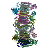
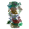
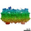
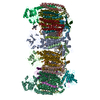
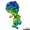
| Title | Cryo-EM structure of hybrid respiratory supercomplex consisting of Mycobacterium tuberculosis complexIII and Mycobacterium smegmatis complexIV in the presence of Q203 | ||||||||||||||||||
 Components Components |
| ||||||||||||||||||
 Keywords Keywords | OXIDOREDUCTASE / Mycobacterium smegmatis / mycobacterium tuberculosis / complexIII / complexIV / electron transport / anti-TB drugs | ||||||||||||||||||
| Function / homology |  Function and homology information Function and homology informationaerobic electron transport chain / cytochrome-c oxidase / quinol-cytochrome-c reductase / quinol-cytochrome-c reductase activity / cytochrome-c oxidase activity / oxidoreductase activity, acting on paired donors, with incorporation or reduction of molecular oxygen / ATP synthesis coupled electron transport / peptidoglycan-based cell wall / respiratory electron transport chain / monooxygenase activity ...aerobic electron transport chain / cytochrome-c oxidase / quinol-cytochrome-c reductase / quinol-cytochrome-c reductase activity / cytochrome-c oxidase activity / oxidoreductase activity, acting on paired donors, with incorporation or reduction of molecular oxygen / ATP synthesis coupled electron transport / peptidoglycan-based cell wall / respiratory electron transport chain / monooxygenase activity / electron transport chain / 2 iron, 2 sulfur cluster binding / oxidoreductase activity / iron ion binding / copper ion binding / heme binding / membrane / metal ion binding / plasma membrane Similarity search - Function | ||||||||||||||||||
| Biological species |  Mycobacterium tuberculosis H37Rv (bacteria)Mycobacterium smegmatis MC2 51 (bacteria)Mycolicibacterium smegmatis MC2 51 (bacteria) Mycobacterium tuberculosis H37Rv (bacteria)Mycobacterium smegmatis MC2 51 (bacteria)Mycolicibacterium smegmatis MC2 51 (bacteria) | ||||||||||||||||||
| Method | ELECTRON MICROSCOPY / single particle reconstruction / cryo EM / Resolution: 2.67 Å | ||||||||||||||||||
 Authors Authors | Zhou, S. / Wang, W. / Gao, Y. / Gong, H. / Rao, Z. | ||||||||||||||||||
| Funding support |  China, 5items China, 5items
| ||||||||||||||||||
 Citation Citation | Journal: Elife / Year: 2021 Title: Structure of cytochrome in complex with Q203 and TB47, two anti-TB drug candidates. Authors: Shan Zhou / Weiwei Wang / Xiaoting Zhou / Yuying Zhang / Yuezheng Lai / Yanting Tang / Jinxu Xu / Dongmei Li / Jianping Lin / Xiaolin Yang / Ting Ran / Hongming Chen / Luke W Guddat / Quan ...Authors: Shan Zhou / Weiwei Wang / Xiaoting Zhou / Yuying Zhang / Yuezheng Lai / Yanting Tang / Jinxu Xu / Dongmei Li / Jianping Lin / Xiaolin Yang / Ting Ran / Hongming Chen / Luke W Guddat / Quan Wang / Yan Gao / Zihe Rao / Hongri Gong /  Abstract: Pathogenic mycobacteria pose a sustained threat to global human health. Recently, cytochrome complexes have gained interest as targets for antibiotic drug development. However, there is currently no ...Pathogenic mycobacteria pose a sustained threat to global human health. Recently, cytochrome complexes have gained interest as targets for antibiotic drug development. However, there is currently no structural information for the cytochrome complex from these pathogenic mycobacteria. Here, we report the structures of cytochrome alone (2.68 Å resolution) and in complex with clinical drug candidates Q203 (2.67 Å resolution) and TB47 (2.93 Å resolution) determined by single-particle cryo-electron microscopy. cytochrome forms a dimeric assembly with endogenous menaquinone/menaquinol bound at the quinone/quinol-binding pockets. We observe Q203 and TB47 bound at the quinol-binding site and stabilized by hydrogen bonds with the side chains of Thr and Glu, residues that are conserved across pathogenic mycobacteria. These high-resolution images provide a basis for the design of new mycobacterial cytochrome inhibitors that could be developed into broad-spectrum drugs to treat mycobacterial infections. | ||||||||||||||||||
| History |
|
- Structure visualization
| Movie |
Movie viewer |
|---|---|
| Structure viewer | Molecule: MolmilJmol/JSmol |
- Downloads & links
-Download
| PDBx/mmCIF format | 7e1w.cif.gz | 948.6 KB | Display | PDBx/mmCIF format |
|---|---|---|---|---|
| PDB format | pdb7e1w.ent.gz | 780.4 KB | Display | PDB format |
| PDBx/mmJSON format | 7e1w.json.gz | Tree view | PDBx/mmJSON format | |
| Others |  Other downloads Other downloads |
-Validation report
| Arichive directory | https://data.pdbj.org/pub/pdb/validation_reports/e1/7e1wftp://data.pdbj.org/pub/pdb/validation_reports/e1/7e1w | HTTPS FTP |
|---|
-Related structure data
| Related structure data |  30944MC  7e1vC  7e1xC M: map data used to model this data C: citing same article ( |
|---|---|
| Similar structure data |


-Links
 PDBj
PDBj


- Assembly
| Deposited unit | 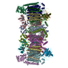
|
|---|---|
| 1 |
|
-Components
-Cytochrome c oxidase subunit ... , 4 types, 8 molecules EQFRGSIU
| #1: Protein | Mass: 38077.465 Da / Num. of mol.: 2 / Source method: isolated from a natural source / Source: (natural) Mycobacterium smegmatis MC2 51 (bacteria) / References: UniProt: A0R057, cytochrome-c oxidase#2: Protein | Mass: 64162.965 Da / Num. of mol.: 2 / Source method: isolated from a natural source Source: (natural) Mycolicibacterium smegmatis MC2 51 (bacteria)#3: Protein | Mass: 22196.883 Da / Num. of mol.: 2 / Source method: isolated from a natural source / Source: (natural) Mycobacterium smegmatis MC2 51 (bacteria) / References: UniProt: A0R049#5: Protein | Mass: 8365.549 Da / Num. of mol.: 2 / Source method: isolated from a natural source / Source: (natural) Mycobacterium smegmatis MC2 51 (bacteria) / References: UniProt: A0R1B6 |
|---|
-Protein , 4 types, 8 molecules HTJVDPMA
| #4: Protein | Mass: 15177.424 Da / Num. of mol.: 2 / Source method: isolated from a natural source / Source: (natural) Mycobacterium smegmatis MC2 51 (bacteria) / References: UniProt: A0R056, cytochrome-c oxidase#6: Protein | Mass: 15910.971 Da / Num. of mol.: 2 / Source method: isolated from a natural source / Source: (natural) Mycobacterium smegmatis MC2 51 (bacteria) / References: UniProt: A0R1B5#7: Protein | Mass: 11329.909 Da / Num. of mol.: 2 / Source method: isolated from a natural source Source: (natural) Mycolicibacterium smegmatis MC2 51 (bacteria)#9: Protein | Mass: 46976.465 Da / Num. of mol.: 2 Source method: isolated from a genetically manipulated source Source: (gene. exp.) Mycobacterium tuberculosis H37Rv (bacteria)Strain: H37Rv / Gene: qcrA, Rv2195, MTCY190.06 Production host: Mycolicibacterium smegmatis MC2 51 (bacteria)References: UniProt: P9WH23 |
|---|
-Cytochrome bc1 complex cytochrome ... , 2 types, 4 molecules NBOC
| #8: Protein | Mass: 61077.062 Da / Num. of mol.: 2 Source method: isolated from a genetically manipulated source Source: (gene. exp.) Mycobacterium tuberculosis H37Rv (bacteria)Strain: H37Rv / Gene: qcrB, Rv2196, MTCY190.07 Production host: Mycolicibacterium smegmatis MC2 51 (bacteria)References: UniProt: P9WP37, quinol-cytochrome-c reductase #10: Protein | Mass: 29152.365 Da / Num. of mol.: 2 Source method: isolated from a genetically manipulated source Source: (gene. exp.) Mycobacterium tuberculosis H37Rv (bacteria)Strain: H37Rv / Gene: qcrC, Rv2194, MTCY190.05 Production host: Mycolicibacterium smegmatis MC2 51 (bacteria)References: UniProt: P9WP35, quinol-cytochrome-c reductase |
|---|
-Non-polymers , 12 types, 60 molecules 


| #11: Chemical | ChemComp-CU /  Mass: 63.546 Da / Num. of mol.: 8 / Source method: obtained synthetically / Formula: Cu Mass: 63.546 Da / Num. of mol.: 8 / Source method: obtained synthetically / Formula: Cu#12: Chemical | ChemComp-CDL /  Mass: 1464.043 Da / Num. of mol.: 17 / Source method: obtained synthetically / Formula: C81H156O17P2 / Comment: phospholipid*YM Mass: 1464.043 Da / Num. of mol.: 17 / Source method: obtained synthetically / Formula: C81H156O17P2 / Comment: phospholipid*YM#13: Chemical | ChemComp-PLM /  Mass: 256.424 Da / Num. of mol.: 4 / Source method: obtained synthetically / Formula: C16H32O2 / Feature type: SUBJECT OF INVESTIGATION Mass: 256.424 Da / Num. of mol.: 4 / Source method: obtained synthetically / Formula: C16H32O2 / Feature type: SUBJECT OF INVESTIGATION#14: Chemical | ChemComp-HEA /  Mass: 852.837 Da / Num. of mol.: 4 / Source method: obtained synthetically / Formula: C49H56FeN4O6 / Feature type: SUBJECT OF INVESTIGATION Mass: 852.837 Da / Num. of mol.: 4 / Source method: obtained synthetically / Formula: C49H56FeN4O6 / Feature type: SUBJECT OF INVESTIGATION#15: Chemical |  Mass: 717.996 Da / Num. of mol.: 2 / Source method: obtained synthetically / Formula: C39H76NO8P / Feature type: SUBJECT OF INVESTIGATION Mass: 717.996 Da / Num. of mol.: 2 / Source method: obtained synthetically / Formula: C39H76NO8P / Feature type: SUBJECT OF INVESTIGATION#16: Chemical | ChemComp-HEM /  Mass: 616.487 Da / Num. of mol.: 4 / Source method: obtained synthetically / Formula: C34H32FeN4O4 / Feature type: SUBJECT OF INVESTIGATION Mass: 616.487 Da / Num. of mol.: 4 / Source method: obtained synthetically / Formula: C34H32FeN4O4 / Feature type: SUBJECT OF INVESTIGATION#17: Chemical | ChemComp-MQ9 /  Mass: 785.233 Da / Num. of mol.: 8 / Source method: obtained synthetically / Formula: C56H80O2 / Feature type: SUBJECT OF INVESTIGATION Mass: 785.233 Da / Num. of mol.: 8 / Source method: obtained synthetically / Formula: C56H80O2 / Feature type: SUBJECT OF INVESTIGATION#18: Chemical |  Mass: 557.006 Da / Num. of mol.: 2 / Source method: obtained synthetically / Formula: C29H28ClF3N4O2 / Feature type: SUBJECT OF INVESTIGATION Mass: 557.006 Da / Num. of mol.: 2 / Source method: obtained synthetically / Formula: C29H28ClF3N4O2 / Feature type: SUBJECT OF INVESTIGATION#19: Chemical |  Mass: 175.820 Da / Num. of mol.: 2 / Source method: obtained synthetically / Formula: Fe2S2 Mass: 175.820 Da / Num. of mol.: 2 / Source method: obtained synthetically / Formula: Fe2S2#20: Chemical | ChemComp-9YF / (  Mass: 853.112 Da / Num. of mol.: 4 / Source method: obtained synthetically / Formula: C44H85O13P / Feature type: SUBJECT OF INVESTIGATION Mass: 853.112 Da / Num. of mol.: 4 / Source method: obtained synthetically / Formula: C44H85O13P / Feature type: SUBJECT OF INVESTIGATION#21: Chemical | ChemComp-HEC /  Mass: 618.503 Da / Num. of mol.: 4 / Source method: obtained synthetically / Formula: C34H34FeN4O4 / Feature type: SUBJECT OF INVESTIGATION Mass: 618.503 Da / Num. of mol.: 4 / Source method: obtained synthetically / Formula: C34H34FeN4O4 / Feature type: SUBJECT OF INVESTIGATION#22: Water | ChemComp-HOH / | Mass: 18.015 Da / Num. of mol.: 1 / Source method: isolated from a natural source / Formula: H2O |
|---|
-Details
| Has ligand of interest | Y |
|---|---|
| Has protein modification | Y |
-Experimental details
-Experiment
| Experiment | Method: ELECTRON MICROSCOPY |
|---|---|
| EM experiment | Aggregation state: PARTICLE / 3D reconstruction method: single particle reconstruction |
- Sample preparation
| Component | Name: hybrid respiratory supercomplex consisting of Mycobacterium tuberculosis complexIII and Mycobacterium smegmatis complexIV in the presence of Q203 Type: COMPLEX / Entity ID: #1-#10 / Source: NATURAL |
|---|---|
| Molecular weight | Value: 0.8 MDa / Experimental value: YES |
| Source (natural) | Organism: Mycolicibacterium smegmatis MC2 51 (bacteria) |
| Buffer solution | pH: 7.4 |
| Specimen | Conc.: 10 mg/ml / Embedding applied: NO / Shadowing applied: NO / Staining applied: NO / Vitrification applied: YES |
| Vitrification | Cryogen name: ETHANE |
- Electron microscopy imaging
| Experimental equipment |  Model: Titan Krios / Image courtesy: FEI Company |
|---|---|
| Microscopy | Model: FEI TITAN KRIOS |
| Electron gun | Electron source:  FIELD EMISSION GUN / Accelerating voltage: 300 kV / Illumination mode: FLOOD BEAM FIELD EMISSION GUN / Accelerating voltage: 300 kV / Illumination mode: FLOOD BEAM |
| Electron lens | Mode: BRIGHT FIELD / Calibrated defocus min: 1200 nm / Calibrated defocus max: 1800 nm |
| Image recording | Electron dose: 60 e/Å2 / Film or detector model: GATAN K3 (6k x 4k) |
- Processing
| Software | Name: PHENIX / Version: 1.16_3549: / Classification: refinement | ||||||||||||||||||||||||
|---|---|---|---|---|---|---|---|---|---|---|---|---|---|---|---|---|---|---|---|---|---|---|---|---|---|
| CTF correction | Type: PHASE FLIPPING AND AMPLITUDE CORRECTION | ||||||||||||||||||||||||
| 3D reconstruction | Resolution: 2.67 Å / Resolution method: FSC 0.143 CUT-OFF / Num. of particles: 106770 / Symmetry type: POINT | ||||||||||||||||||||||||
| Refine LS restraints |
|
