 Movie
Movie Controller


+ Open data

- Basic information
| Entry | Database: PDB / ID: 7dsc | ||||||
|---|---|---|---|---|---|---|---|
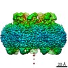
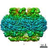
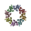
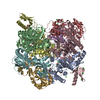
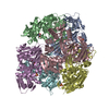
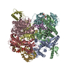
| Title | CALHM1 open state with disordered CTH | ||||||
 Components Components | Calcium homeostasis modulator 1 | ||||||
 Keywords Keywords | MEMBRANE PROTEIN / channel | ||||||
| Function / homology | Calcium homeostasis modulator family / Calcium homeostasis modulator / ATP export / voltage-gated calcium channel activity / plasma membrane / Calcium homeostasis modulator 1 Function and homology information Function and homology information | ||||||
| Biological species |  | ||||||
| Method | ELECTRON MICROSCOPY / single particle reconstruction / cryo EM / Resolution: 3.5 Å | ||||||
 Authors Authors | Ren, Y. / Yang, X. / Shen, Y.Q. | ||||||
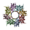
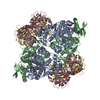
 Citation Citation | Journal: J Biol Chem / Year: 2022 Title: Cryo-EM structure of the heptameric calcium homeostasis modulator 1 channel. Authors: Yue Ren / Yang Li / Yaojie Wang / Tianlei Wen / Xuhang Lu / Shenghai Chang / Xing Zhang / Yuequan Shen / Xue Yang /  Abstract: Calcium homeostasis modulator 1 (CALHM1) is a voltage- and Ca-gated ATP channel that plays an important role in neuronal signaling. However, as the previously reported CALHM structures are all in the ...Calcium homeostasis modulator 1 (CALHM1) is a voltage- and Ca-gated ATP channel that plays an important role in neuronal signaling. However, as the previously reported CALHM structures are all in the ATP-conducting state, the gating mechanism of ATP permeation is still elusive. Here, we report cryo-EM reconstructions of two Danio rerio CALHM1 heptamers with ordered or flexible long C-terminal helices at resolutions of 3.2 Å and 2.9 Å, respectively, and one D. rerio CALHM1 octamer with flexible long C-terminal helices at a resolution of 3.5 Å. Structural analysis shows that the heptameric CALHM1s are in an ATP-nonconducting state with a central pore diameter of approximately 6.6 Å. Compared with those inside the octameric CALHM1, the N-helix inside the heptameric CALHM1 is in the "down" position to avoid steric clashing with the adjacent TM1 helix. Molecular dynamics simulations show that as the N-helix moves from the "down" position to the "up" position, the pore size of ATP molecule permeation increases significantly. Our results provide important information for elucidating the mechanism of ATP molecule permeation in the CALHM1 channel. | ||||||
| History |
|
- Structure visualization
| Movie |
Movie viewer |
|---|---|
| Structure viewer | Molecule: MolmilJmol/JSmol |
- Downloads & links
-Download
| PDBx/mmCIF format | 7dsc.cif.gz | 281.8 KB | Display | PDBx/mmCIF format |
|---|---|---|---|---|
| PDB format | pdb7dsc.ent.gz | 220.5 KB | Display | PDB format |
| PDBx/mmJSON format | 7dsc.json.gz | Tree view | PDBx/mmJSON format | |
| Others |  Other downloads Other downloads |
-Validation report
| Summary document | 7dsc_validation.pdf.gz | 816 KB | Display | wwPDB validaton report |
|---|---|---|---|---|
| Full document | 7dsc_full_validation.pdf.gz | 823.3 KB | Display | |
| Data in XML | 7dsc_validation.xml.gz | 43 KB | Display | |
| Data in CIF | 7dsc_validation.cif.gz | 54.9 KB | Display | |
| Arichive directory | https://data.pdbj.org/pub/pdb/validation_reports/ds/7dscftp://data.pdbj.org/pub/pdb/validation_reports/ds/7dsc | HTTPS FTP |
-Related structure data
| Related structure data | 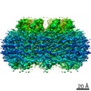 30830MC  7dsdC  7dseC M: map data used to model this data C: citing same article ( |
|---|---|
| Similar structure data |
-Links
 PDBj
PDBj- Assembly
| Deposited unit | 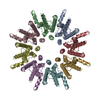
|
|---|---|
| 1 |
|
-Components
| #1: Protein | Mass: 41213.645 Da / Num. of mol.: 8 Source method: isolated from a genetically manipulated source Source: (gene. exp.)  Homo sapiens (human) / References: UniProt: E7F2J4 Homo sapiens (human) / References: UniProt: E7F2J4#2: Sugar | ChemComp-NAG /   Type: D-saccharide, beta linking / Mass: 221.208 Da / Num. of mol.: 8 / Source method: obtained synthetically / Formula: C8H15NO6 Type: D-saccharide, beta linking / Mass: 221.208 Da / Num. of mol.: 8 / Source method: obtained synthetically / Formula: C8H15NO6Has ligand of interest | N | Has protein modification | Y | |
|---|
-Experimental details
-Experiment
| Experiment | Method: ELECTRON MICROSCOPY |
|---|---|
| EM experiment | Aggregation state: 2D ARRAY / 3D reconstruction method: single particle reconstruction |
- Sample preparation
| Component | Name: CALHM1 / Type: ORGANELLE OR CELLULAR COMPONENT / Entity ID: #1 / Source: RECOMBINANT |
|---|---|
| Molecular weight | Experimental value: NO |
| Source (natural) | Organism: |
| Source (recombinant) | Organism: Homo sapiens (human) / Cell: HEK-293S GnTI- |
| Buffer solution | pH: 7.5 |
| Specimen | Conc.: 10 mg/ml / Embedding applied: NO / Shadowing applied: NO / Staining applied: NO / Vitrification applied: YES |
| Specimen support | Grid material: GOLD / Grid type: Quantifoil |
| Vitrification | Instrument: FEI VITROBOT MARK IV / Cryogen name: ETHANE / Humidity: 100 % / Chamber temperature: 281 K |
- Electron microscopy imaging
| Experimental equipment |  Model: Titan Krios / Image courtesy: FEI Company |
|---|---|
| Microscopy | Model: FEI TITAN KRIOS |
| Electron gun | Electron source:  FIELD EMISSION GUN / Accelerating voltage: 300 kV / Illumination mode: FLOOD BEAM FIELD EMISSION GUN / Accelerating voltage: 300 kV / Illumination mode: FLOOD BEAM |
| Electron lens | Mode: BRIGHT FIELD |
| Specimen holder | Cryogen: NITROGEN |
| Image recording | Electron dose: 50 e/Å2 / Detector mode: COUNTING / Film or detector model: GATAN K2 SUMMIT (4k x 4k) |
- Processing
| EM software | Name: SerialEM / Category: image acquisition |
|---|---|
| CTF correction | Type: NONE |
| 3D reconstruction | Resolution: 3.5 Å / Resolution method: FSC 0.143 CUT-OFF / Num. of particles: 44894 / Symmetry type: POINT |
