+ Open data
- Basic information
| Entry | Database: PDB / ID: 7c2t | |||||||||||||||||||||
|---|---|---|---|---|---|---|---|---|---|---|---|---|---|---|---|---|---|---|---|---|---|---|
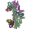
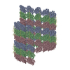
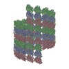
| Title | Helical reconstruction of Zika virus complexed with Fab C10 | |||||||||||||||||||||
 Components Components |
| |||||||||||||||||||||
 Keywords Keywords | VIRUS / antibody / neutralization | |||||||||||||||||||||
| Function / homology |  Function and homology information Function and homology informationflavivirin / symbiont-mediated suppression of host JAK-STAT cascade via inhibition of host TYK2 activity / symbiont-mediated suppression of host JAK-STAT cascade via inhibition of STAT2 activity / symbiont-mediated suppression of host JAK-STAT cascade via inhibition of STAT1 activity / negative regulation of innate immune response / ribonucleoside triphosphate phosphatase activity / viral capsid / double-stranded RNA binding / nucleoside-triphosphate phosphatase / 4 iron, 4 sulfur cluster binding ...flavivirin / symbiont-mediated suppression of host JAK-STAT cascade via inhibition of host TYK2 activity / symbiont-mediated suppression of host JAK-STAT cascade via inhibition of STAT2 activity / symbiont-mediated suppression of host JAK-STAT cascade via inhibition of STAT1 activity / negative regulation of innate immune response / ribonucleoside triphosphate phosphatase activity / viral capsid / double-stranded RNA binding / nucleoside-triphosphate phosphatase / 4 iron, 4 sulfur cluster binding / clathrin-dependent endocytosis of virus by host cell / mRNA (guanine-N7)-methyltransferase / methyltransferase cap1 / molecular adaptor activity / methyltransferase cap1 activity / mRNA 5'-cap (guanine-N7-)-methyltransferase activity / RNA helicase activity / protein dimerization activity / symbiont-mediated suppression of host innate immune response / host cell perinuclear region of cytoplasm / host cell endoplasmic reticulum membrane / RNA helicase / symbiont-mediated suppression of host type I interferon-mediated signaling pathway / symbiont-mediated activation of host autophagy / serine-type endopeptidase activity / RNA-directed RNA polymerase / viral RNA genome replication / RNA-directed RNA polymerase activity / fusion of virus membrane with host endosome membrane / viral envelope / centrosome / symbiont entry into host cell / lipid binding / GTP binding / virion attachment to host cell / host cell nucleus / virion membrane / structural molecule activity / ATP hydrolysis activity / proteolysis / extracellular region / ATP binding / metal ion binding / membrane Similarity search - Function | |||||||||||||||||||||
| Biological species |  Homo sapiens (human) Homo sapiens (human)  Zika virus Zika virus | |||||||||||||||||||||
| Method | ELECTRON MICROSCOPY / helical reconstruction / cryo EM / Resolution: 9.4 Å | |||||||||||||||||||||
 Authors Authors | Morrone, S. / Chew, S.V. / Lim, X.N. / Ng, T.S. / Kostyuchenko, V.A. / Zhang, S. / Lok, S.M. | |||||||||||||||||||||
| Funding support |  Singapore, 2items Singapore, 2items
| |||||||||||||||||||||
 Citation Citation |  Journal: Nat Commun / Year: 2020 Journal: Nat Commun / Year: 2020Title: High flavivirus structural plasticity demonstrated by a non-spherical morphological variant. Authors: Seamus R Morrone / Valerie S Y Chew / Xin-Ni Lim / Thiam-Seng Ng / Victor A Kostyuchenko / Shuijun Zhang / Melissa Wirawan / Pau-Ling Chew / Jaime Lee / Joanne L Tan / Jiaqi Wang / Ter Yong ...Authors: Seamus R Morrone / Valerie S Y Chew / Xin-Ni Lim / Thiam-Seng Ng / Victor A Kostyuchenko / Shuijun Zhang / Melissa Wirawan / Pau-Ling Chew / Jaime Lee / Joanne L Tan / Jiaqi Wang / Ter Yong Tan / Jian Shi / Gavin Screaton / Marc C Morais / Shee-Mei Lok /    Abstract: Previous flavivirus (dengue and Zika viruses) studies showed largely spherical particles either with smooth or bumpy surfaces. Here, we demonstrate flavivirus particles have high structural ...Previous flavivirus (dengue and Zika viruses) studies showed largely spherical particles either with smooth or bumpy surfaces. Here, we demonstrate flavivirus particles have high structural plasticity by the induction of a non-spherical morphology at elevated temperatures: the club-shaped particle (clubSP), which contains a cylindrical tail and a disc-like head. Complex formation of DENV and ZIKV with Fab C10 stabilize the viruses allowing cryoEM structural determination to ~10 Å resolution. The caterpillar-shaped (catSP) Fab C10:ZIKV complex shows Fabs locking the E protein raft structure containing three E dimers. However, compared to the original spherical structure, the rafts have rotated relative to each other. The helical tail structure of Fab C10:DENV3 clubSP showed although the Fab locked an E protein dimer, the dimers have shifted laterally. Morphological diversity, including clubSP and the previously identified bumpy and smooth-surfaced spherical particles, may help flavivirus survival and immune evasion. | |||||||||||||||||||||
| History |
|
- Structure visualization
| Movie |
 Movie viewer Movie viewer |
|---|---|
| Structure viewer | Molecule:  Molmil Molmil Jmol/JSmol Jmol/JSmol |
- Downloads & links
- Download
| PDBx/mmCIF format |  7c2t.cif.gz 7c2t.cif.gz | 67.2 KB | Display |  PDBx/mmCIF format PDBx/mmCIF format |
|---|---|---|---|---|
| PDB format |  pdb7c2t.ent.gz pdb7c2t.ent.gz | 40.1 KB | Display |  PDB format PDB format |
| PDBx/mmJSON format |  7c2t.json.gz 7c2t.json.gz | Tree view |  PDBx/mmJSON format PDBx/mmJSON format | |
| Others |  Other downloads Other downloads |
-Validation report
| Summary document |  7c2t_validation.pdf.gz 7c2t_validation.pdf.gz | 917.8 KB | Display |  wwPDB validaton report wwPDB validaton report |
|---|---|---|---|---|
| Full document |  7c2t_full_validation.pdf.gz 7c2t_full_validation.pdf.gz | 917.4 KB | Display | |
| Data in XML |  7c2t_validation.xml.gz 7c2t_validation.xml.gz | 25.4 KB | Display | |
| Data in CIF |  7c2t_validation.cif.gz 7c2t_validation.cif.gz | 37.5 KB | Display | |
| Arichive directory |  https://data.pdbj.org/pub/pdb/validation_reports/c2/7c2t https://data.pdbj.org/pub/pdb/validation_reports/c2/7c2t ftp://data.pdbj.org/pub/pdb/validation_reports/c2/7c2t ftp://data.pdbj.org/pub/pdb/validation_reports/c2/7c2t | HTTPS FTP |
-Related structure data
| Related structure data |  30279MC  7c2sC M: map data used to model this data C: citing same article ( |
|---|---|
| Similar structure data |
- Links
Links
- Assembly
| Deposited unit | 
|
|---|---|
| 1 | x 30
|
- Components
| #1: Protein | Mass: 54444.051 Da / Num. of mol.: 2 / Source method: isolated from a natural source / Source: (natural)   Zika virus / References: UniProt: A0A2D1AHP1, UniProt: A0A024B7W1*PLUS Zika virus / References: UniProt: A0A2D1AHP1, UniProt: A0A024B7W1*PLUS#2: Protein | Mass: 8496.883 Da / Num. of mol.: 2 / Source method: isolated from a natural source / Source: (natural)   Zika virus / References: UniProt: A0A2D1AQS6, UniProt: A0A024B7W1*PLUS Zika virus / References: UniProt: A0A2D1AQS6, UniProt: A0A024B7W1*PLUS#3: Antibody | Mass: 14487.058 Da / Num. of mol.: 2 Source method: isolated from a genetically manipulated source Source: (gene. exp.)  Homo sapiens (human) / Cell line (production host): HEK293T / Production host: Homo sapiens (human) / Cell line (production host): HEK293T / Production host:  Homo sapiens (human) Homo sapiens (human)#4: Antibody | Mass: 11298.362 Da / Num. of mol.: 2 Source method: isolated from a genetically manipulated source Source: (gene. exp.)  Homo sapiens (human) / Cell line (production host): HEK293T / Production host: Homo sapiens (human) / Cell line (production host): HEK293T / Production host:  Homo sapiens (human) Homo sapiens (human)Has protein modification | N | |
|---|
-Experimental details
-Experiment
| Experiment | Method: ELECTRON MICROSCOPY |
|---|---|
| EM experiment | Aggregation state: FILAMENT / 3D reconstruction method: helical reconstruction |
- Sample preparation
| Component |
| ||||||||||||||||||||||||
|---|---|---|---|---|---|---|---|---|---|---|---|---|---|---|---|---|---|---|---|---|---|---|---|---|---|
| Source (natural) |
| ||||||||||||||||||||||||
| Source (recombinant) | Organism:  Homo sapiens (human) / Cell: HEK293-6E Homo sapiens (human) / Cell: HEK293-6E | ||||||||||||||||||||||||
| Buffer solution | pH: 8 | ||||||||||||||||||||||||
| Specimen | Embedding applied: NO / Shadowing applied: NO / Staining applied: NO / Vitrification applied: YES | ||||||||||||||||||||||||
| Vitrification | Cryogen name: ETHANE |
- Electron microscopy imaging
| Experimental equipment |  Model: Titan Krios / Image courtesy: FEI Company |
|---|---|
| Microscopy | Model: FEI TITAN KRIOS |
| Electron gun | Electron source:  FIELD EMISSION GUN / Accelerating voltage: 300 kV / Illumination mode: FLOOD BEAM FIELD EMISSION GUN / Accelerating voltage: 300 kV / Illumination mode: FLOOD BEAM |
| Electron lens | Mode: BRIGHT FIELD |
| Image recording | Electron dose: 38 e/Å2 / Film or detector model: FEI FALCON II (4k x 4k) |
- Processing
| Software | Name: PHENIX / Version: 1.10.1_2155: / Classification: refinement | ||||||||||||||||||||||||
|---|---|---|---|---|---|---|---|---|---|---|---|---|---|---|---|---|---|---|---|---|---|---|---|---|---|
| EM software | Name: PHENIX / Category: model refinement | ||||||||||||||||||||||||
| CTF correction | Type: PHASE FLIPPING AND AMPLITUDE CORRECTION | ||||||||||||||||||||||||
| Helical symmerty | Angular rotation/subunit: 24.1 ° / Axial rise/subunit: 8.6 Å / Axial symmetry: C1 | ||||||||||||||||||||||||
| 3D reconstruction | Resolution: 9.4 Å / Resolution method: FSC 0.143 CUT-OFF / Num. of particles: 3406 / Symmetry type: HELICAL | ||||||||||||||||||||||||
| Refine LS restraints |
|
 Movie
Movie Controller


 PDBj
PDBj


