 Movie
Movie Controller


+ Open data

- Basic information
| Entry | Database: PDB / ID: 7bzw | ||||||
|---|---|---|---|---|---|---|---|
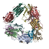
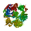
| Title | Structure of Hsp21 | ||||||
 Components Components | Heat shock protein 21, chloroplastic | ||||||
 Keywords Keywords | CHAPERONE / small heat shock protein | ||||||
| Function / homology |  Function and homology information Function and homology informationchloroplast nucleoid / chloroplast organization / response to light stimulus / protein folding chaperone complex / response to heat / regulation of DNA-templated transcription Similarity search - Function | ||||||
| Biological species |  | ||||||
| Method | ELECTRON MICROSCOPY / single particle reconstruction / cryo EM / Resolution: 4.6 Å | ||||||
 Authors Authors | Lau, W.C.Y. | ||||||
| Funding support |  Hong Kong, 1items Hong Kong, 1items
| ||||||
 Citation Citation | Journal: Nat Commun / Year: 2021 Title: Structural basis of substrate recognition and thermal protection by a small heat shock protein. Authors: Chuanyang Yu / Stephen King Pong Leung / Wenxin Zhang / Louis Tung Faat Lai / Ying Ki Chan / Man Chit Wong / Samir Benlekbir / Yong Cui / Liwen Jiang / Wilson Chun Yu Lau /  Abstract: Small heat shock proteins (sHsps) bind unfolding proteins, thereby playing a pivotal role in the maintenance of proteostasis in virtually all living organisms. Structural elucidation of sHsp- ...Small heat shock proteins (sHsps) bind unfolding proteins, thereby playing a pivotal role in the maintenance of proteostasis in virtually all living organisms. Structural elucidation of sHsp-substrate complexes has been hampered by the transient and heterogeneous nature of their interactions, and the precise mechanisms underlying substrate recognition, promiscuity, and chaperone activity of sHsps remain unclear. Here we show the formation of a stable complex between Arabidopsis thaliana plastid sHsp, Hsp21, and its natural substrate 1-deoxy-D-xylulose 5-phosphate synthase (DXPS) under heat stress, and report cryo-electron microscopy structures of Hsp21, DXPS and Hsp21-DXPS complex at near-atomic resolution. Monomeric Hsp21 binds across the dimer interface of DXPS and engages in multivalent interactions by recognizing highly dynamic structural elements in DXPS. Hsp21 partly unfolds its central α-crystallin domain to facilitate binding of DXPS, which preserves a native-like structure. This mode of interaction suggests a mechanism of sHsps anti-aggregation activity towards a broad range of substrates. | ||||||
| History |
|
- Structure visualization
| Movie |
Movie viewer |
|---|---|
| Structure viewer | Molecule: MolmilJmol/JSmol |
- Downloads & links
-Download
| PDBx/mmCIF format | 7bzw.cif.gz | 234.5 KB | Display | PDBx/mmCIF format |
|---|---|---|---|---|
| PDB format | pdb7bzw.ent.gz | Display | PDB format | |
| PDBx/mmJSON format | 7bzw.json.gz | Tree view | PDBx/mmJSON format | |
| Others |  Other downloads Other downloads |
-Validation report
| Arichive directory | https://data.pdbj.org/pub/pdb/validation_reports/bz/7bzwftp://data.pdbj.org/pub/pdb/validation_reports/bz/7bzw | HTTPS FTP |
|---|
-Related structure data
| Related structure data | 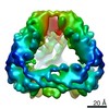 30261MC  7bzxC  7bzyC M: map data used to model this data C: citing same article ( |
|---|---|
| Similar structure data |


-Links
 PDBj
PDBj


- Assembly
| Deposited unit | 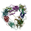
|
|---|---|
| 1 |
|
-Components
| #1: Protein | Mass: 23654.545 Da / Num. of mol.: 12 Source method: isolated from a genetically manipulated source Source: (gene. exp.)  |
|---|
-Experimental details
-Experiment
| Experiment | Method: ELECTRON MICROSCOPY |
|---|---|
| EM experiment | Aggregation state: PARTICLE / 3D reconstruction method: single particle reconstruction |
- Sample preparation
| Component | Name: Hsp21 / Type: COMPLEX / Entity ID: all / Source: RECOMBINANT |
|---|---|
| Molecular weight | Value: 0.25 MDa / Experimental value: NO |
| Source (natural) | Organism: |
| Source (recombinant) | Organism: |
| Buffer solution | pH: 8 |
| Specimen | Embedding applied: NO / Shadowing applied: NO / Staining applied: NO / Vitrification applied: YES |
| Vitrification | Cryogen name: ETHANE |
- Electron microscopy imaging
| Experimental equipment |  Model: Titan Krios / Image courtesy: FEI Company |
|---|---|
| Microscopy | Model: FEI TITAN KRIOS |
| Electron gun | Electron source:  FIELD EMISSION GUN / Accelerating voltage: 300 kV / Illumination mode: FLOOD BEAM FIELD EMISSION GUN / Accelerating voltage: 300 kV / Illumination mode: FLOOD BEAM |
| Electron lens | Mode: BRIGHT FIELD |
| Image recording | Electron dose: 45 e/Å2 / Detector mode: COUNTING / Film or detector model: FEI FALCON III (4k x 4k) |
- Processing
| CTF correction | Type: PHASE FLIPPING AND AMPLITUDE CORRECTION |
|---|---|
| Symmetry | Point symmetry: T (tetrahedral) |
| 3D reconstruction | Resolution: 4.6 Å / Resolution method: FSC 0.143 CUT-OFF / Num. of particles: 104126 / Symmetry type: POINT |
