 Movie
Movie Controller


+ Open data

- Basic information
| Entry | Database: PDB / ID: 7z6w | |||||||||
|---|---|---|---|---|---|---|---|---|---|---|
| Title | Complex of E. coli LolA and lipoprotein | |||||||||
 Components Components | Outer-membrane lipoprotein carrier protein | |||||||||
 Keywords Keywords | TRANSPORT PROTEIN / Lipoprotein trafficking / PROTEIN TRANSPORT | |||||||||
| Function / homology | Outer membrane lipoprotein carrier protein LolA, Proteobacteria / Outer membrane lipoprotein carrier protein LolA / Outer membrane lipoprotein carrier protein LolA-like / Lipoprotein localisation LolA/LolB/LppX / lipoprotein localization to outer membrane / lipoprotein transport / outer membrane-bounded periplasmic space / Chem-IG7 / Outer-membrane lipoprotein carrier protein Function and homology information Function and homology information | |||||||||
| Biological species |  | |||||||||
| Method |  X-RAY DIFFRACTION / SYNCHROTRON / MOLECULAR REPLACEMENT / Resolution: 1.84 Å X-RAY DIFFRACTION / SYNCHROTRON / MOLECULAR REPLACEMENT / Resolution: 1.84 Å | |||||||||
 Authors Authors | Kaplan, E. / Greene, N.P. / Koronakis, V. | |||||||||
| Funding support |  United Kingdom, 2items United Kingdom, 2items
| |||||||||
 Citation Citation | Journal: Proc.Natl.Acad.Sci.USA / Year: 2022 Title: Structural basis of lipoprotein recognition by the bacterial Lol trafficking chaperone LolA. Authors: Kaplan, E. / Greene, N.P. / Jepson, A.E. / Koronakis, V. | |||||||||
| History |
|
- Structure visualization
| Structure viewer | Molecule: MolmilJmol/JSmol |
|---|
- Downloads & links
-Download
| PDBx/mmCIF format | 7z6w.cif.gz | 56.2 KB | Display | PDBx/mmCIF format |
|---|---|---|---|---|
| PDB format | pdb7z6w.ent.gz | 38 KB | Display | PDB format |
| PDBx/mmJSON format | 7z6w.json.gz | Tree view | PDBx/mmJSON format | |
| Others |  Other downloads Other downloads |
-Validation report
| Summary document | 7z6w_validation.pdf.gz | 745.9 KB | Display | wwPDB validaton report |
|---|---|---|---|---|
| Full document | 7z6w_full_validation.pdf.gz | 748.8 KB | Display | |
| Data in XML | 7z6w_validation.xml.gz | 10.7 KB | Display | |
| Data in CIF | 7z6w_validation.cif.gz | 14.1 KB | Display | |
| Arichive directory | https://data.pdbj.org/pub/pdb/validation_reports/z6/7z6wftp://data.pdbj.org/pub/pdb/validation_reports/z6/7z6w | HTTPS FTP |
-Related structure data
| Related structure data | 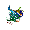 7z6xC 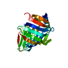 1ua8S S: Starting model for refinement C: citing same article ( |
|---|---|
| Similar structure data |
-Links
 PDBj
PDBj- Assembly
| Deposited unit | 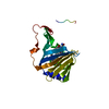
| ||||||||
|---|---|---|---|---|---|---|---|---|---|
| 1 |
| ||||||||
| Unit cell |
|
-Components
| #1: Protein | Mass: 21529.570 Da / Num. of mol.: 1 Source method: isolated from a genetically manipulated source Source: (gene. exp.) |
|---|---|
| #2: Chemical | ChemComp-IG7 / [(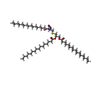  Mass: 909.478 Da / Num. of mol.: 1 / Source method: obtained synthetically / Formula: C54H104N2O6S / Feature type: SUBJECT OF INVESTIGATION Mass: 909.478 Da / Num. of mol.: 1 / Source method: obtained synthetically / Formula: C54H104N2O6S / Feature type: SUBJECT OF INVESTIGATION |
| #3: Water | ChemComp-HOH /  Mass: 18.015 Da / Num. of mol.: 74 / Source method: isolated from a natural source / Formula: H2O Mass: 18.015 Da / Num. of mol.: 74 / Source method: isolated from a natural source / Formula: H2O |
| Has ligand of interest | Y |
-Experimental details
-Experiment
| Experiment | Method: X-RAY DIFFRACTION / Number of used crystals: 1 |
|---|
- Sample preparation
| Crystal | Density Matthews: 2.29 Å3/Da / Density % sol: 46.19 % |
|---|---|
| Crystal grow | Temperature: 288.15 K / Method: vapor diffusion, sitting drop / Details: 2.1 M DL-Malic acid pH 6.0 |
-Data collection
| Diffraction | Mean temperature: 100 K / Serial crystal experiment: N | ||||||||||||||||||||||||||||||
|---|---|---|---|---|---|---|---|---|---|---|---|---|---|---|---|---|---|---|---|---|---|---|---|---|---|---|---|---|---|---|---|
| Diffraction source | Source: SYNCHROTRON / Site: Diamond / Beamline: I04-1 / Wavelength: 0.91587 Å | ||||||||||||||||||||||||||||||
| Detector | Type: DECTRIS PILATUS 6M-F / Detector: PIXEL / Date: Apr 8, 2019 | ||||||||||||||||||||||||||||||
| Radiation | Protocol: SINGLE WAVELENGTH / Monochromatic (M) / Laue (L): M / Scattering type: x-ray | ||||||||||||||||||||||||||||||
| Radiation wavelength | Wavelength: 0.91587 Å / Relative weight: 1 | ||||||||||||||||||||||||||||||
| Reflection | Resolution: 1.84→64.33 Å / Num. obs: 17042 / % possible obs: 100 % / Redundancy: 12.1 % / CC1/2: 0.999 / Rmerge(I) obs: 0.124 / Rpim(I) all: 0.037 / Rrim(I) all: 0.13 / Net I/σ(I): 14.3 / Num. measured all: 207055 / Scaling rejects: 12 | ||||||||||||||||||||||||||||||
| Reflection shell | Diffraction-ID: 1
|
- Processing
| Software |
| ||||||||||||||||||||||||||||||||||||||||||||||||||||||||||||
|---|---|---|---|---|---|---|---|---|---|---|---|---|---|---|---|---|---|---|---|---|---|---|---|---|---|---|---|---|---|---|---|---|---|---|---|---|---|---|---|---|---|---|---|---|---|---|---|---|---|---|---|---|---|---|---|---|---|---|---|---|---|
| Refinement | Method to determine structure: MOLECULAR REPLACEMENT Starting model: 1UA8 Resolution: 1.84→64.33 Å / Cor.coef. Fo:Fc: 0.962 / Cor.coef. Fo:Fc free: 0.954 / SU B: 3.393 / SU ML: 0.098 / Cross valid method: THROUGHOUT / σ(F): 0 / ESU R: 0.139 / ESU R Free: 0.128 / Stereochemistry target values: MAXIMUM LIKELIHOOD Details: HYDROGENS HAVE BEEN ADDED IN THE RIDING POSITIONS U VALUES : REFINED INDIVIDUALLY
| ||||||||||||||||||||||||||||||||||||||||||||||||||||||||||||
| Solvent computation | Ion probe radii: 0.8 Å / Shrinkage radii: 0.8 Å / VDW probe radii: 1.2 Å / Solvent model: MASK | ||||||||||||||||||||||||||||||||||||||||||||||||||||||||||||
| Displacement parameters | Biso max: 91.33 Å2 / Biso mean: 29.02 Å2 / Biso min: 14.69 Å2
| ||||||||||||||||||||||||||||||||||||||||||||||||||||||||||||
| Refinement step | Cycle: final / Resolution: 1.84→64.33 Å
| ||||||||||||||||||||||||||||||||||||||||||||||||||||||||||||
| Refine LS restraints |
| ||||||||||||||||||||||||||||||||||||||||||||||||||||||||||||
| LS refinement shell | Resolution: 1.84→1.888 Å / Rfactor Rfree error: 0 / Total num. of bins used: 20
|
