 Movie
Movie Controller

[English] 日本語
 Yorodumi
- PDB-7wmt: Crystal structure of small molecule 13 bound to human Nicotinamid... -
+ Open data

- Basic information
| Entry | Database: PDB / ID: 7wmt | ||||||
|---|---|---|---|---|---|---|---|
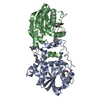
| Title | Crystal structure of small molecule 13 bound to human Nicotinamide N-methyltransferase | ||||||
 Components Components | Nicotinamide N-methyltransferase | ||||||
 Keywords Keywords | TRANSFERASE / Nicotinamide N-methyltransferase | ||||||
| Function / homology |  Function and homology information Function and homology informationpyridine N-methyltransferase activity / nicotinamide N-methyltransferase / positive regulation of protein deacetylation / nicotinamide N-methyltransferase activity / Metabolism of ingested SeMet, Sec, MeSec into H2Se / nicotinamide metabolic process / nicotinate metabolic process / Methylation / NAD+ catabolic process / Nicotinate metabolism ...pyridine N-methyltransferase activity / nicotinamide N-methyltransferase / positive regulation of protein deacetylation / nicotinamide N-methyltransferase activity / Metabolism of ingested SeMet, Sec, MeSec into H2Se / nicotinamide metabolic process / nicotinate metabolic process / Methylation / NAD+ catabolic process / Nicotinate metabolism / positive regulation of gluconeogenesis / methylation / cytosol Similarity search - Function | ||||||
| Biological species |  Homo sapiens (human) Homo sapiens (human) | ||||||
| Method |  X-RAY DIFFRACTION / MOLECULAR REPLACEMENT / molecular replacement / Resolution: 1.77 Å X-RAY DIFFRACTION / MOLECULAR REPLACEMENT / molecular replacement / Resolution: 1.77 Å | ||||||
 Authors Authors | Yoshida, S. / Uehara, S. / Kondo, N. / Takahashi, Y. / Yamamoto, S. / Kameda, A. / Kawagoe, S. / Inoue, N. / Yamada, M. / Yoshimura, N. / Tachibana, Y. | ||||||
| Funding support | 1items
| ||||||
 Citation Citation | Journal: J.Med.Chem. / Year: 2022 Title: Peptide-to-Small Molecule: A Pharmacophore-Guided Small Molecule Lead Generation Strategy from High-Affinity Macrocyclic Peptides. Authors: Yoshida, S. / Uehara, S. / Kondo, N. / Takahashi, Y. / Yamamoto, S. / Kameda, A. / Kawagoe, S. / Inoue, N. / Yamada, M. / Yoshimura, N. / Tachibana, Y. | ||||||
| History |
|
- Structure visualization
| Structure viewer | Molecule: MolmilJmol/JSmol |
|---|
- Downloads & links
-Download
| PDBx/mmCIF format | 7wmt.cif.gz | 67.2 KB | Display | PDBx/mmCIF format |
|---|---|---|---|---|
| PDB format | pdb7wmt.ent.gz | 46.3 KB | Display | PDB format |
| PDBx/mmJSON format | 7wmt.json.gz | Tree view | PDBx/mmJSON format | |
| Others |  Other downloads Other downloads |
-Validation report
| Arichive directory | https://data.pdbj.org/pub/pdb/validation_reports/wm/7wmtftp://data.pdbj.org/pub/pdb/validation_reports/wm/7wmt | HTTPS FTP |
|---|
-Related structure data
-Links
 PDBj
PDBj- Assembly
| Deposited unit | 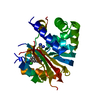
| ||||||||
|---|---|---|---|---|---|---|---|---|---|
| 1 |
| ||||||||
| Unit cell |
|
-Components
| #1: Protein | Mass: 27683.770 Da / Num. of mol.: 1 Source method: isolated from a genetically manipulated source Source: (gene. exp.) Homo sapiens (human) / Gene: NNMT / Production host:  References: UniProt: P40261, nicotinamide N-methyltransferase |
|---|---|
| #2: Chemical | ChemComp-1IV / [(  Mass: 564.080 Da / Num. of mol.: 1 / Source method: obtained synthetically / Formula: C32H30ClN7O / Feature type: SUBJECT OF INVESTIGATION Mass: 564.080 Da / Num. of mol.: 1 / Source method: obtained synthetically / Formula: C32H30ClN7O / Feature type: SUBJECT OF INVESTIGATION |
| #3: Water | ChemComp-HOH /  Mass: 18.015 Da / Num. of mol.: 202 / Source method: isolated from a natural source / Formula: H2O Mass: 18.015 Da / Num. of mol.: 202 / Source method: isolated from a natural source / Formula: H2O |
| Has ligand of interest | Y |
-Experimental details
-Experiment
| Experiment | Method: X-RAY DIFFRACTION / Number of used crystals: 1 |
|---|
- Sample preparation
| Crystal | Density Matthews: 2.75 Å3/Da / Density % sol: 55.23 % |
|---|---|
| Crystal grow | Temperature: 277 K / Method: vapor diffusion, sitting drop Details: 0.04 M Potassium phosphate monobasic, 16 % w/v PEG 8000, 20 % v/v Glycerol |
-Data collection
| Diffraction | Mean temperature: 100 K / Serial crystal experiment: N | |||||||||||||||||||||||||||||||||||||||||||||||||||||||||||||||||||||||||||||||||||||||||||||||||||
|---|---|---|---|---|---|---|---|---|---|---|---|---|---|---|---|---|---|---|---|---|---|---|---|---|---|---|---|---|---|---|---|---|---|---|---|---|---|---|---|---|---|---|---|---|---|---|---|---|---|---|---|---|---|---|---|---|---|---|---|---|---|---|---|---|---|---|---|---|---|---|---|---|---|---|---|---|---|---|---|---|---|---|---|---|---|---|---|---|---|---|---|---|---|---|---|---|---|---|---|---|
| Diffraction source | Source: ROTATING ANODE / Type: RIGAKU FR-E+ SUPERBRIGHT / Wavelength: 1.54178 Å | |||||||||||||||||||||||||||||||||||||||||||||||||||||||||||||||||||||||||||||||||||||||||||||||||||
| Detector | Type: RIGAKU RAXIS VII / Detector: IMAGE PLATE / Date: Feb 25, 2019 | |||||||||||||||||||||||||||||||||||||||||||||||||||||||||||||||||||||||||||||||||||||||||||||||||||
| Radiation | Protocol: SINGLE WAVELENGTH / Monochromatic (M) / Laue (L): M / Scattering type: x-ray | |||||||||||||||||||||||||||||||||||||||||||||||||||||||||||||||||||||||||||||||||||||||||||||||||||
| Radiation wavelength | Wavelength: 1.54178 Å / Relative weight: 1 | |||||||||||||||||||||||||||||||||||||||||||||||||||||||||||||||||||||||||||||||||||||||||||||||||||
| Reflection | Resolution: 1.77→54.26 Å / Num. obs: 28583 / % possible obs: 99.4 % / Redundancy: 5 % / Rmerge(I) obs: 0.036 / Rpim(I) all: 0.017 / Rrim(I) all: 0.04 / Χ2: 1.082 / Net I/σ(I): 28.1 | |||||||||||||||||||||||||||||||||||||||||||||||||||||||||||||||||||||||||||||||||||||||||||||||||||
| Reflection shell | Diffraction-ID: 1
|
-Phasing
| Phasing | Method: molecular replacement | ||||||
|---|---|---|---|---|---|---|---|
| Phasing MR | R rigid body: 0.35
|
- Processing
| Software |
| |||||||||||||||||||||||||||||||||||||||||||||
|---|---|---|---|---|---|---|---|---|---|---|---|---|---|---|---|---|---|---|---|---|---|---|---|---|---|---|---|---|---|---|---|---|---|---|---|---|---|---|---|---|---|---|---|---|---|---|
| Refinement | Method to determine structure: MOLECULAR REPLACEMENT / Resolution: 1.77→54.26 Å / Cor.coef. Fo:Fc: 0.974 / Cor.coef. Fo:Fc free: 0.963 / SU B: 1.927 / SU ML: 0.062 / SU R Cruickshank DPI: 0.0926 / Cross valid method: THROUGHOUT / σ(F): 0 / ESU R: 0.093 / ESU R Free: 0.095 / Stereochemistry target values: MAXIMUM LIKELIHOOD Details: HYDROGENS HAVE BEEN USED IF PRESENT IN THE INPUT U VALUES : REFINED INDIVIDUALLY
| |||||||||||||||||||||||||||||||||||||||||||||
| Solvent computation | Ion probe radii: 0.8 Å / Shrinkage radii: 0.8 Å / VDW probe radii: 1.2 Å / Solvent model: BABINET MODEL WITH MASK | |||||||||||||||||||||||||||||||||||||||||||||
| Displacement parameters | Biso max: 114.48 Å2 / Biso mean: 43.953 Å2 / Biso min: 22.53 Å2
| |||||||||||||||||||||||||||||||||||||||||||||
| Refinement step | Cycle: final / Resolution: 1.77→54.26 Å
| |||||||||||||||||||||||||||||||||||||||||||||
| Refine LS restraints |
| |||||||||||||||||||||||||||||||||||||||||||||
| LS refinement shell | Resolution: 1.772→1.818 Å / Rfactor Rfree error: 0 / Total num. of bins used: 20
|
