 Movie
Movie Controller

[English] 日本語
 Yorodumi
- PDB-7vyp: The structure of GdmN complex with the natural tetrahedral interm... -
+ Open data

- Basic information
| Entry | Database: PDB / ID: 7vyp | ||||||||||||||||||||||||
|---|---|---|---|---|---|---|---|---|---|---|---|---|---|---|---|---|---|---|---|---|---|---|---|---|---|
| Title | The structure of GdmN complex with the natural tetrahedral intermediate, carbamoylated derivative, and AMP | ||||||||||||||||||||||||
 Components Components | GdmN | ||||||||||||||||||||||||
 Keywords Keywords | TRANSFERASE / Carbamoylation / Ansamycins antibiotics / Homodimer | ||||||||||||||||||||||||
| Function / homology |  Function and homology information Function and homology informationbiosynthetic process / catalytic activity / ATP binding / metal ion binding Similarity search - Function | ||||||||||||||||||||||||
| Biological species |  Streptomyces hygroscopicus (bacteria) Streptomyces hygroscopicus (bacteria) | ||||||||||||||||||||||||
| Method |  X-RAY DIFFRACTION / SYNCHROTRON / MOLECULAR REPLACEMENT / Resolution: 2.88 Å X-RAY DIFFRACTION / SYNCHROTRON / MOLECULAR REPLACEMENT / Resolution: 2.88 Å | ||||||||||||||||||||||||
 Authors Authors | Wei, J. / Zheng, J. / Zhou, J. / Kang, Q. / Bai, L. | ||||||||||||||||||||||||
| Funding support | 7items
| ||||||||||||||||||||||||
 Citation Citation | Journal: Nat Commun / Year: 2022 Title: Endowing homodimeric carbamoyltransferase GdmN with iterative functions through structural characterization and mechanistic studies. Authors: Wei, J. / Zhang, X. / Zhou, Y. / Cheng, X. / Lin, Z. / Tang, M. / Zheng, J. / Wang, B. / Kang, Q. / Bai, L. | ||||||||||||||||||||||||
| History |
|
- Structure visualization
| Structure viewer | Molecule: MolmilJmol/JSmol |
|---|
- Downloads & links
-Download
| PDBx/mmCIF format | 7vyp.cif.gz | 336.7 KB | Display | PDBx/mmCIF format |
|---|---|---|---|---|
| PDB format | pdb7vyp.ent.gz | 217.9 KB | Display | PDB format |
| PDBx/mmJSON format | 7vyp.json.gz | Tree view | PDBx/mmJSON format | |
| Others |  Other downloads Other downloads |
-Validation report
| Summary document | 7vyp_validation.pdf.gz | 1.3 MB | Display | wwPDB validaton report |
|---|---|---|---|---|
| Full document | 7vyp_full_validation.pdf.gz | 1.3 MB | Display | |
| Data in XML | 7vyp_validation.xml.gz | 49.4 KB | Display | |
| Data in CIF | 7vyp_validation.cif.gz | 66.8 KB | Display | |
| Arichive directory | https://data.pdbj.org/pub/pdb/validation_reports/vy/7vypftp://data.pdbj.org/pub/pdb/validation_reports/vy/7vyp | HTTPS FTP |
-Related structure data
| Related structure data |  7vx0C  7vyjC  7vyoC  7vznC 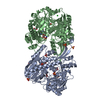 7vzqC 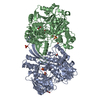 7vzuC  7vzyC  7vzzC  3venS S: Starting model for refinement C: citing same article ( |
|---|---|
| Similar structure data |
-Links
 PDBj
PDBj


- Assembly
| Deposited unit | 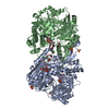
| ||||||||||||
|---|---|---|---|---|---|---|---|---|---|---|---|---|---|
| 1 |
| ||||||||||||
| Unit cell |
|
-Components
-Protein , 1 types, 2 molecules AB
| #1: Protein | Mass: 74365.609 Da / Num. of mol.: 2 Source method: isolated from a genetically manipulated source Source: (gene. exp.) Streptomyces hygroscopicus (bacteria) / Gene: gdmN / Production host: |
|---|
-Non-polymers , 8 types, 94 molecules 

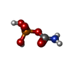

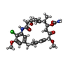


| #2: Chemical |  Mass: 55.845 Da / Num. of mol.: 2 / Source method: obtained synthetically / Formula: Fe Mass: 55.845 Da / Num. of mol.: 2 / Source method: obtained synthetically / Formula: Fe#3: Chemical | ChemComp-SO4 /  Mass: 96.063 Da / Num. of mol.: 8 / Source method: obtained synthetically / Formula: SO4 Mass: 96.063 Da / Num. of mol.: 8 / Source method: obtained synthetically / Formula: SO4#4: Chemical | ChemComp-CP / |  Mass: 141.020 Da / Num. of mol.: 1 / Source method: obtained synthetically / Formula: CH4NO5P Mass: 141.020 Da / Num. of mol.: 1 / Source method: obtained synthetically / Formula: CH4NO5P#5: Chemical | ChemComp-EDO /  Mass: 62.068 Da / Num. of mol.: 10 / Source method: obtained synthetically / Formula: C2H6O2 Mass: 62.068 Da / Num. of mol.: 10 / Source method: obtained synthetically / Formula: C2H6O2#6: Chemical | ChemComp-82Z / [( |  Mass: 535.029 Da / Num. of mol.: 1 / Source method: obtained synthetically / Formula: C27H35ClN2O7 / Feature type: SUBJECT OF INVESTIGATION Mass: 535.029 Da / Num. of mol.: 1 / Source method: obtained synthetically / Formula: C27H35ClN2O7 / Feature type: SUBJECT OF INVESTIGATION#7: Chemical | ChemComp-AMP / |  Mass: 347.221 Da / Num. of mol.: 1 / Source method: isolated from a natural source / Formula: C10H14N5O7P / Feature type: SUBJECT OF INVESTIGATION / Comment: AMP*YM Mass: 347.221 Da / Num. of mol.: 1 / Source method: isolated from a natural source / Formula: C10H14N5O7P / Feature type: SUBJECT OF INVESTIGATION / Comment: AMP*YM#8: Chemical | ChemComp-8CW / [( |  Mass: 882.250 Da / Num. of mol.: 1 / Source method: obtained synthetically / Formula: C37H49ClN7O14P / Feature type: SUBJECT OF INVESTIGATION Mass: 882.250 Da / Num. of mol.: 1 / Source method: obtained synthetically / Formula: C37H49ClN7O14P / Feature type: SUBJECT OF INVESTIGATION#9: Water | ChemComp-HOH / | Mass: 18.015 Da / Num. of mol.: 70 / Source method: isolated from a natural source / Formula: H2O |
|---|
-Details
| Has ligand of interest | Y |
|---|
-Experimental details
-Experiment
| Experiment | Method: X-RAY DIFFRACTION / Number of used crystals: 1 |
|---|
- Sample preparation
| Crystal | Density Matthews: 2.79 Å3/Da / Density % sol: 55.94 % |
|---|---|
| Crystal grow | Temperature: 293.15 K / Method: vapor diffusion, sitting drop Details: Lithium sulfate monohydrate, Polyethylene glycol 3,350, Tris |
-Data collection
| Diffraction | Mean temperature: 100 K / Serial crystal experiment: N |
|---|---|
| Diffraction source | Source: SYNCHROTRON / Site: SSRF  / Beamline: BL18U1 / Wavelength: 0.97853 Å / Beamline: BL18U1 / Wavelength: 0.97853 Å |
| Detector | Type: DECTRIS PILATUS3 6M / Detector: PIXEL / Date: May 2, 2019 |
| Radiation | Protocol: SINGLE WAVELENGTH / Monochromatic (M) / Laue (L): M / Scattering type: x-ray |
| Radiation wavelength | Wavelength: 0.97853 Å / Relative weight: 1 |
| Reflection | Resolution: 2.88→50 Å / Num. obs: 38176 / % possible obs: 100 % / Observed criterion σ(F): 0 / Observed criterion σ(I): -3 / Redundancy: 9.8 % / Biso Wilson estimate: 49.6 Å2 / CC1/2: 0.989 / CC star: 0.997 / Rmerge(I) obs: 0.129 / Rpim(I) all: 0.043 / Rrim(I) all: 0.136 / Rsym value: 0.129 / Χ2: 0.907 / Net I/σ(I): 21.167 |
| Reflection shell | Resolution: 2.88→2.93 Å / Num. unique obs: 1880 / CC1/2: 0.973 |
- Processing
| Software |
| ||||||||||||||||||||||||||||||||||||||||||||||||||||||||||||||||||||||||||||||||||||||||||||||||||
|---|---|---|---|---|---|---|---|---|---|---|---|---|---|---|---|---|---|---|---|---|---|---|---|---|---|---|---|---|---|---|---|---|---|---|---|---|---|---|---|---|---|---|---|---|---|---|---|---|---|---|---|---|---|---|---|---|---|---|---|---|---|---|---|---|---|---|---|---|---|---|---|---|---|---|---|---|---|---|---|---|---|---|---|---|---|---|---|---|---|---|---|---|---|---|---|---|---|---|---|
| Refinement | Method to determine structure: MOLECULAR REPLACEMENT Starting model: 3VEN Resolution: 2.88→27.76 Å / SU ML: 0.2955 / Cross valid method: FREE R-VALUE / σ(F): 1.34 / Phase error: 22.1538 Stereochemistry target values: GeoStd + Monomer Library + CDL v1.2
| ||||||||||||||||||||||||||||||||||||||||||||||||||||||||||||||||||||||||||||||||||||||||||||||||||
| Solvent computation | Shrinkage radii: 0.9 Å / VDW probe radii: 1.11 Å / Solvent model: FLAT BULK SOLVENT MODEL | ||||||||||||||||||||||||||||||||||||||||||||||||||||||||||||||||||||||||||||||||||||||||||||||||||
| Displacement parameters | Biso mean: 43.76 Å2 | ||||||||||||||||||||||||||||||||||||||||||||||||||||||||||||||||||||||||||||||||||||||||||||||||||
| Refinement step | Cycle: LAST / Resolution: 2.88→27.76 Å
| ||||||||||||||||||||||||||||||||||||||||||||||||||||||||||||||||||||||||||||||||||||||||||||||||||
| Refine LS restraints |
| ||||||||||||||||||||||||||||||||||||||||||||||||||||||||||||||||||||||||||||||||||||||||||||||||||
| LS refinement shell |
|
