 Movie
Movie Controller


+ Open data

- Basic information
| Entry | Database: PDB / ID: 7uf8 | |||||||||
|---|---|---|---|---|---|---|---|---|---|---|
| Title | Structure of CtdP in complex with penicimutamide E and NADP+ | |||||||||
 Components Components | CtdP | |||||||||
 Keywords Keywords | OXIDOREDUCTASE / Diels-Alderase / oxidioreductase / NmrA-like | |||||||||
| Function / homology | Penicimutamide E / NADP NICOTINAMIDE-ADENINE-DINUCLEOTIDE PHOSPHATE Function and homology information Function and homology information | |||||||||
| Biological species |  Penicillium citrinum (fungus) Penicillium citrinum (fungus) | |||||||||
| Method |  X-RAY DIFFRACTION / SYNCHROTRON / SAD / Resolution: 2.5 Å X-RAY DIFFRACTION / SYNCHROTRON / SAD / Resolution: 2.5 Å | |||||||||
 Authors Authors | Rivera, S. / Liu, Z. / Newmister, S.A. / Gao, X. / Sherman, D.H. | |||||||||
| Funding support |  United States, 2items United States, 2items
| |||||||||
 Citation Citation | Journal: Nat.Chem. / Year: 2023 Title: An NmrA-like enzyme-catalysed redox-mediated Diels-Alder cycloaddition with anti-selectivity. Authors: Liu, Z. / Rivera, S. / Newmister, S.A. / Sanders, J.N. / Nie, Q. / Liu, S. / Zhao, F. / Ferrara, J.D. / Shih, H.W. / Patil, S. / Xu, W. / Miller, M.D. / Phillips, G.N. / Houk, K.N. / Sherman, D.H. / Gao, X. | |||||||||
| History |
|
- Structure visualization
| Structure viewer | Molecule: MolmilJmol/JSmol |
|---|
- Downloads & links
-Download
| PDBx/mmCIF format | 7uf8.cif.gz | 601.4 KB | Display | PDBx/mmCIF format |
|---|---|---|---|---|
| PDB format | pdb7uf8.ent.gz | 414.4 KB | Display | PDB format |
| PDBx/mmJSON format | 7uf8.json.gz | Tree view | PDBx/mmJSON format | |
| Others |  Other downloads Other downloads |
-Validation report
| Summary document | 7uf8_validation.pdf.gz | 2.5 MB | Display | wwPDB validaton report |
|---|---|---|---|---|
| Full document | 7uf8_full_validation.pdf.gz | 2.5 MB | Display | |
| Data in XML | 7uf8_validation.xml.gz | 50.4 KB | Display | |
| Data in CIF | 7uf8_validation.cif.gz | 67.2 KB | Display | |
| Arichive directory | https://data.pdbj.org/pub/pdb/validation_reports/uf/7uf8ftp://data.pdbj.org/pub/pdb/validation_reports/uf/7uf8 | HTTPS FTP |
-Related structure data
| Similar structure data |
|---|
-Links
 PDBj
PDBj- Assembly
| Deposited unit | 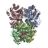
| ||||||||||||
|---|---|---|---|---|---|---|---|---|---|---|---|---|---|
| 1 |
| ||||||||||||
| Unit cell |
|
-Components
| #1: Protein | Mass: 39013.984 Da / Num. of mol.: 4 Source method: isolated from a genetically manipulated source Source: (gene. exp.) Penicillium citrinum (fungus) / Gene: ctdP / Production host:  #2: Chemical | ChemComp-NAP /   Mass: 743.405 Da / Num. of mol.: 4 / Source method: obtained synthetically / Formula: C21H28N7O17P3 / Feature type: SUBJECT OF INVESTIGATION Mass: 743.405 Da / Num. of mol.: 4 / Source method: obtained synthetically / Formula: C21H28N7O17P3 / Feature type: SUBJECT OF INVESTIGATION#3: Chemical |   Mass: 335.443 Da / Num. of mol.: 3 / Source method: obtained synthetically / Formula: C21H25N3O / Feature type: SUBJECT OF INVESTIGATION Mass: 335.443 Da / Num. of mol.: 3 / Source method: obtained synthetically / Formula: C21H25N3O / Feature type: SUBJECT OF INVESTIGATION#4: Chemical |   Mass: 62.068 Da / Num. of mol.: 2 / Source method: obtained synthetically / Formula: C2H6O2 Mass: 62.068 Da / Num. of mol.: 2 / Source method: obtained synthetically / Formula: C2H6O2#5: Water | ChemComp-HOH / |  Mass: 18.015 Da / Num. of mol.: 115 / Source method: isolated from a natural source / Formula: H2O Mass: 18.015 Da / Num. of mol.: 115 / Source method: isolated from a natural source / Formula: H2OHas ligand of interest | Y | |
|---|
-Experimental details
-Experiment
| Experiment | Method: X-RAY DIFFRACTION / Number of used crystals: 1 |
|---|
- Sample preparation
| Crystal | Density Matthews: 2.5 Å3/Da / Density % sol: 50.9 % |
|---|---|
| Crystal grow | Temperature: 293 K / Method: vapor diffusion, sitting drop / pH: 6.75 Details: 0.1 M BisTris pH 6.75, 25% PEG monomethyl ether 2000, 0.1 M CaCl2 |
-Data collection
| Diffraction | Mean temperature: 100 K / Serial crystal experiment: N |
|---|---|
| Diffraction source | Source: SYNCHROTRON / Site: APS / Beamline: 23-ID-B / Wavelength: 0.9795 Å |
| Detector | Type: DECTRIS EIGER X 16M / Detector: PIXEL / Date: Jun 11, 2021 |
| Radiation | Protocol: SINGLE WAVELENGTH / Monochromatic (M) / Laue (L): M / Scattering type: x-ray |
| Radiation wavelength | Wavelength: 0.9795 Å / Relative weight: 1 |
| Reflection | Resolution: 2.5→48.36 Å / Num. obs: 55814 / % possible obs: 99.8 % / Redundancy: 21.13 % / Biso Wilson estimate: 60.06 Å2 / CC1/2: 0.999 / Rrim(I) all: 0.174 / Net I/σ(I): 17.8 |
| Reflection shell | Resolution: 2.5→2.56 Å / Rmerge(I) obs: 2.79 / Mean I/σ(I) obs: 1.7 / Num. unique obs: 16814 / CC1/2: 0.826 / Rrim(I) all: 2.85 / % possible all: 98.8 |
- Processing
| Software |
| |||||||||||||||||||||||||||||||||||||||||||||||||||||||||||||||||||||||||||||||||||||||||||||||||||||||||
|---|---|---|---|---|---|---|---|---|---|---|---|---|---|---|---|---|---|---|---|---|---|---|---|---|---|---|---|---|---|---|---|---|---|---|---|---|---|---|---|---|---|---|---|---|---|---|---|---|---|---|---|---|---|---|---|---|---|---|---|---|---|---|---|---|---|---|---|---|---|---|---|---|---|---|---|---|---|---|---|---|---|---|---|---|---|---|---|---|---|---|---|---|---|---|---|---|---|---|---|---|---|---|---|---|---|---|
| Refinement | Method to determine structure: SAD / Resolution: 2.5→48.36 Å / SU ML: 0.3985 / Cross valid method: FREE R-VALUE / σ(F): 1.34 / Phase error: 31.7196 Stereochemistry target values: GeoStd + Monomer Library + CDL v1.2
| |||||||||||||||||||||||||||||||||||||||||||||||||||||||||||||||||||||||||||||||||||||||||||||||||||||||||
| Solvent computation | Shrinkage radii: 0.9 Å / VDW probe radii: 1.11 Å / Solvent model: FLAT BULK SOLVENT MODEL | |||||||||||||||||||||||||||||||||||||||||||||||||||||||||||||||||||||||||||||||||||||||||||||||||||||||||
| Displacement parameters | Biso mean: 72.89 Å2 | |||||||||||||||||||||||||||||||||||||||||||||||||||||||||||||||||||||||||||||||||||||||||||||||||||||||||
| Refinement step | Cycle: LAST / Resolution: 2.5→48.36 Å
| |||||||||||||||||||||||||||||||||||||||||||||||||||||||||||||||||||||||||||||||||||||||||||||||||||||||||
| Refine LS restraints |
| |||||||||||||||||||||||||||||||||||||||||||||||||||||||||||||||||||||||||||||||||||||||||||||||||||||||||
| LS refinement shell |
|
