 Movie
Movie Controller


+ Open data

- Basic information
| Entry | Database: PDB / ID: 7taa | ||||||
|---|---|---|---|---|---|---|---|
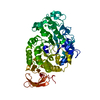
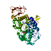
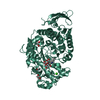
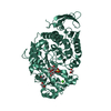
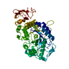
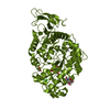
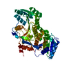
| Title | FAMILY 13 ALPHA AMYLASE IN COMPLEX WITH ACARBOSE | ||||||
 Components Components | TAKA AMYLASE | ||||||
 Keywords Keywords | HYDROLASE / GLYCOSYL HYDROLASE / TAKA / AMYLASE / ACARBOSE | ||||||
| Function / homology |  Function and homology information Function and homology informationcell wall-bounded periplasmic space / hyphal septin band / spitzenkorper / alpha-amylase / fungal-type cell wall / cell septum / alpha-amylase activity / carbohydrate catabolic process / calcium ion binding / extracellular region Similarity search - Function | ||||||
| Biological species |  | ||||||
| Method |  X-RAY DIFFRACTION / NATIVE STRUCTURE / Resolution: 1.98 Å X-RAY DIFFRACTION / NATIVE STRUCTURE / Resolution: 1.98 Å | ||||||
 Authors Authors | Davies, G.J. / Brzozowski, A.M. | ||||||
 Citation Citation | Journal: Biochemistry / Year: 1997 Title: Structure of the Aspergillus oryzae alpha-amylase complexed with the inhibitor acarbose at 2.0 A resolution. Authors: Brzozowski, A.M. / Davies, G.J. | ||||||
| History |
|
- Structure visualization
| Structure viewer | Molecule: MolmilJmol/JSmol |
|---|
- Downloads & links
-Download
| PDBx/mmCIF format | 7taa.cif.gz | 117 KB | Display | PDBx/mmCIF format |
|---|---|---|---|---|
| PDB format | pdb7taa.ent.gz | 89.1 KB | Display | PDB format |
| PDBx/mmJSON format | 7taa.json.gz | Tree view | PDBx/mmJSON format | |
| Others |  Other downloads Other downloads |
-Validation report
| Arichive directory | https://data.pdbj.org/pub/pdb/validation_reports/ta/7taaftp://data.pdbj.org/pub/pdb/validation_reports/ta/7taa | HTTPS FTP |
|---|
-Related structure data
| Similar structure data |
|---|


-Links
 PDBj
PDBj

- Assembly
| Deposited unit | 
| ||||||||
|---|---|---|---|---|---|---|---|---|---|
| 1 |
| ||||||||
| Unit cell |
|
-Components
| #1: Protein | Mass: 52525.973 Da / Num. of mol.: 1 / Source method: isolated from a natural source / Source: (natural) References: UniProt: P10529, UniProt: P0C1B3*PLUS, alpha-amylase |
|---|---|
| #2: Sugar | ChemComp-ABC /   Type: saccharide / Mass: 937.887 Da / Num. of mol.: 1 Type: saccharide / Mass: 937.887 Da / Num. of mol.: 1Source method: isolated from a genetically manipulated source Formula: C37H63NO26 |
| #3: Chemical | ChemComp-CA /   Mass: 40.078 Da / Num. of mol.: 1 / Source method: obtained synthetically / Formula: Ca Mass: 40.078 Da / Num. of mol.: 1 / Source method: obtained synthetically / Formula: Ca |
| #4: Water | ChemComp-HOH /  Mass: 18.015 Da / Num. of mol.: 465 / Source method: isolated from a natural source / Formula: H2O Mass: 18.015 Da / Num. of mol.: 465 / Source method: isolated from a natural source / Formula: H2O |
| Has protein modification | Y |
-Experimental details
-Experiment
| Experiment | Method: X-RAY DIFFRACTION / Number of used crystals: 1 |
|---|
- Sample preparation
| Crystal | Density Matthews: 2.19 Å3/Da / Density % sol: 44 % | ||||||||||||||||||||||||||||||||||||||||||
|---|---|---|---|---|---|---|---|---|---|---|---|---|---|---|---|---|---|---|---|---|---|---|---|---|---|---|---|---|---|---|---|---|---|---|---|---|---|---|---|---|---|---|---|
| Crystal grow | Method: vapor diffusion, hanging drop / pH: 7.5 Details: 18% MM PEG 5000, 5MM CALCIUM CHLORIDE, 0.1M HEPES BUFFER, PH 7.5. METHOD: HANGING DROP VAPOUR DIFFUSION. THE NATIVE CRYSTALS WERE SOAKED FOR 6 HOURS IN STABILIZING SOLUTION CONTAINING 15 MM ...Details: 18% MM PEG 5000, 5MM CALCIUM CHLORIDE, 0.1M HEPES BUFFER, PH 7.5. METHOD: HANGING DROP VAPOUR DIFFUSION. THE NATIVE CRYSTALS WERE SOAKED FOR 6 HOURS IN STABILIZING SOLUTION CONTAINING 15 MM OF ACARBOSE., vapor diffusion - hanging drop | ||||||||||||||||||||||||||||||||||||||||||
| Crystal | *PLUS | ||||||||||||||||||||||||||||||||||||||||||
| Crystal grow | *PLUS Temperature: 18 ℃ / Method: vapor diffusion, sitting drop | ||||||||||||||||||||||||||||||||||||||||||
| Components of the solutions | *PLUS
|
-Data collection
| Diffraction | Mean temperature: 120 K |
|---|---|
| Diffraction source | Source: ROTATING ANODE / Type: RIGAKU / Wavelength: 1.5418 |
| Detector | Type: MARRESEARCH / Detector: IMAGE PLATE / Date: 1995 / Details: LONG MIRRORS |
| Radiation | Monochromatic (M) / Laue (L): M / Scattering type: x-ray |
| Radiation wavelength | Wavelength: 1.5418 Å / Relative weight: 1 |
| Reflection | Resolution: 1.98→20 Å / Num. obs: 63935 / % possible obs: 97 % / Redundancy: 3.7 % / Rmerge(I) obs: 0.088 / Net I/σ(I): 12.6 |
| Reflection shell | Resolution: 1.98→2.09 Å / Redundancy: 3.7 % / Rmerge(I) obs: 0.291 / Mean I/σ(I) obs: 5.1 / % possible all: 94.2 |
| Reflection | *PLUS Num. obs: 31062 / Num. measured all: 113411 |
| Reflection shell | *PLUS % possible obs: 94.2 % |
- Processing
| Software |
| ||||||||||||||||||||||||||||||||||||||||||||||||||||||||||||||||||||||||||||||||||||
|---|---|---|---|---|---|---|---|---|---|---|---|---|---|---|---|---|---|---|---|---|---|---|---|---|---|---|---|---|---|---|---|---|---|---|---|---|---|---|---|---|---|---|---|---|---|---|---|---|---|---|---|---|---|---|---|---|---|---|---|---|---|---|---|---|---|---|---|---|---|---|---|---|---|---|---|---|---|---|---|---|---|---|---|---|---|
| Refinement | Method to determine structure: NATIVE STRUCTURE / Resolution: 1.98→20 Å / Cross valid method: FREE R
| ||||||||||||||||||||||||||||||||||||||||||||||||||||||||||||||||||||||||||||||||||||
| Refinement step | Cycle: LAST / Resolution: 1.98→20 Å
| ||||||||||||||||||||||||||||||||||||||||||||||||||||||||||||||||||||||||||||||||||||
| Refine LS restraints |
| ||||||||||||||||||||||||||||||||||||||||||||||||||||||||||||||||||||||||||||||||||||
| LS refinement shell | Resolution: 1.98→2.07 Å / Total num. of bins used: 20
| ||||||||||||||||||||||||||||||||||||||||||||||||||||||||||||||||||||||||||||||||||||
| Software | *PLUS Name: REFMAC / Classification: refinement | ||||||||||||||||||||||||||||||||||||||||||||||||||||||||||||||||||||||||||||||||||||
| Refinement | *PLUS Rfactor obs: 0.159 | ||||||||||||||||||||||||||||||||||||||||||||||||||||||||||||||||||||||||||||||||||||
| Solvent computation | *PLUS | ||||||||||||||||||||||||||||||||||||||||||||||||||||||||||||||||||||||||||||||||||||
| Displacement parameters | *PLUS |
