 Movie
Movie Controller


+ Open data

- Basic information
| Entry | Database: PDB / ID: 7s7a | |||||||||
|---|---|---|---|---|---|---|---|---|---|---|
| Title | Crystal structure of CDK2 liganded with compound EF3019 | |||||||||
 Components Components | Cyclin-dependent kinase 2 | |||||||||
 Keywords Keywords | CELL CYCLE / allosteric inhibitor / drug development / kinase | |||||||||
| Function / homology | Replication initiator protein RctB, central region / RctB, helix turn helix domain / Vibrionales, replication initiator protein RctB, central region / RctB helix turn helix domain / Chem-8FI / Uncharacterized protein Function and homology information Function and homology information | |||||||||
| Biological species |  Homo sapiens (human) Homo sapiens (human) | |||||||||
| Method |  X-RAY DIFFRACTION / SYNCHROTRON / MOLECULAR REPLACEMENT / Resolution: 1.7 Å X-RAY DIFFRACTION / SYNCHROTRON / MOLECULAR REPLACEMENT / Resolution: 1.7 Å | |||||||||
 Authors Authors | Sun, L. / Schonbrunn, E. | |||||||||
| Funding support |  United States, 2items United States, 2items
| |||||||||
 Citation Citation | Journal: J.Med.Chem. / Year: 2023 Title: Screening through Lead Optimization of High Affinity, Allosteric Cyclin-Dependent Kinase 2 (CDK2) Inhibitors as Male Contraceptives That Reduce Sperm Counts in Mice. Authors: Faber, E.B. / Wang, N. / John, K. / Sun, L. / Wong, H.L. / Burban, D. / Francis, R. / Tian, D. / Hong, K.H. / Yang, A. / Wang, L. / Elsaid, M. / Khalid, H. / Levinson, N.M. / Schonbrunn, E. ...Authors: Faber, E.B. / Wang, N. / John, K. / Sun, L. / Wong, H.L. / Burban, D. / Francis, R. / Tian, D. / Hong, K.H. / Yang, A. / Wang, L. / Elsaid, M. / Khalid, H. / Levinson, N.M. / Schonbrunn, E. / Hawkinson, J.E. / Georg, G.I. | |||||||||
| History |
|
- Structure visualization
| Structure viewer | Molecule: MolmilJmol/JSmol |
|---|
- Downloads & links
-Download
| PDBx/mmCIF format | 7s7a.cif.gz | 73.5 KB | Display | PDBx/mmCIF format |
|---|---|---|---|---|
| PDB format | pdb7s7a.ent.gz | 51.8 KB | Display | PDB format |
| PDBx/mmJSON format | 7s7a.json.gz | Tree view | PDBx/mmJSON format | |
| Others |  Other downloads Other downloads |
-Validation report
| Summary document | 7s7a_validation.pdf.gz | 706.6 KB | Display | wwPDB validaton report |
|---|---|---|---|---|
| Full document | 7s7a_full_validation.pdf.gz | 711.2 KB | Display | |
| Data in XML | 7s7a_validation.xml.gz | 13.2 KB | Display | |
| Data in CIF | 7s7a_validation.cif.gz | 18.1 KB | Display | |
| Arichive directory | https://data.pdbj.org/pub/pdb/validation_reports/s7/7s7aftp://data.pdbj.org/pub/pdb/validation_reports/s7/7s7a | HTTPS FTP |
-Related structure data
| Related structure data | 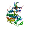 7rweC 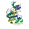 7rxoC 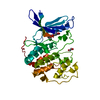 7s4tC 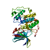 7s85C 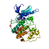 4kd1S S: Starting model for refinement C: citing same article ( |
|---|---|
| Similar structure data |
-Links
 PDBj
PDBj
- Assembly
| Deposited unit | 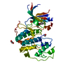
| ||||||||
|---|---|---|---|---|---|---|---|---|---|
| 1 |
| ||||||||
| Unit cell |
|
-Components
| #1: Protein | Mass: 33976.488 Da / Num. of mol.: 1 Source method: isolated from a genetically manipulated source Source: (gene. exp.) Homo sapiens (human) / Gene: CDK2, CDKN2 / Production host:  | ||||||
|---|---|---|---|---|---|---|---|
| #2: Chemical |   Mass: 62.068 Da / Num. of mol.: 3 / Source method: obtained synthetically / Formula: C2H6O2 Mass: 62.068 Da / Num. of mol.: 3 / Source method: obtained synthetically / Formula: C2H6O2#3: Chemical | ChemComp-8FI / | 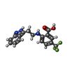  Mass: 349.307 Da / Num. of mol.: 1 / Source method: isolated from a natural source / Formula: C17H14F3N3O2 / Feature type: SUBJECT OF INVESTIGATION Mass: 349.307 Da / Num. of mol.: 1 / Source method: isolated from a natural source / Formula: C17H14F3N3O2 / Feature type: SUBJECT OF INVESTIGATION#4: Water | ChemComp-HOH / |  Mass: 18.015 Da / Num. of mol.: 115 / Source method: isolated from a natural source / Formula: H2O Mass: 18.015 Da / Num. of mol.: 115 / Source method: isolated from a natural source / Formula: H2OHas ligand of interest | Y | |
-Experimental details
-Experiment
| Experiment | Method: X-RAY DIFFRACTION / Number of used crystals: 1 |
|---|
- Sample preparation
| Crystal | Density Matthews: 2.08 Å3/Da / Density % sol: 40.8 % |
|---|---|
| Crystal grow | Temperature: 291 K / Method: vapor diffusion, hanging drop Details: 4.5mg/mL CDK2 protein crystalized under 25mM Na/K phosphate, 25mM Hepes Na, pH 7.5, 5% v/v PEG3350 was soaked overnight with 5mM EF3109 in 50mM Hepes Na, pH 7.5, 10% v/v PEG3350 |
-Data collection
| Diffraction | Mean temperature: 93 K / Serial crystal experiment: N | ||||||||||||||||||||||||||||||||||||||||||||||||||||||||||||||||||||||||||||||||||||
|---|---|---|---|---|---|---|---|---|---|---|---|---|---|---|---|---|---|---|---|---|---|---|---|---|---|---|---|---|---|---|---|---|---|---|---|---|---|---|---|---|---|---|---|---|---|---|---|---|---|---|---|---|---|---|---|---|---|---|---|---|---|---|---|---|---|---|---|---|---|---|---|---|---|---|---|---|---|---|---|---|---|---|---|---|---|
| Diffraction source | Source: SYNCHROTRON / Site: APS / Beamline: 23-ID-D / Wavelength: 1.0332 Å | ||||||||||||||||||||||||||||||||||||||||||||||||||||||||||||||||||||||||||||||||||||
| Detector | Type: DECTRIS PILATUS 6M / Detector: PIXEL / Date: Mar 10, 2020 | ||||||||||||||||||||||||||||||||||||||||||||||||||||||||||||||||||||||||||||||||||||
| Radiation | Protocol: SINGLE WAVELENGTH / Monochromatic (M) / Laue (L): M / Scattering type: x-ray | ||||||||||||||||||||||||||||||||||||||||||||||||||||||||||||||||||||||||||||||||||||
| Radiation wavelength | Wavelength: 1.0332 Å / Relative weight: 1 | ||||||||||||||||||||||||||||||||||||||||||||||||||||||||||||||||||||||||||||||||||||
| Reflection | Resolution: 1.7→27.77 Å / Num. obs: 31791 / % possible obs: 99.69 % / Redundancy: 3.3 % / Biso Wilson estimate: 28.56 Å2 / Rrim(I) all: 0.045 / Net I/σ(I): 21.14 | ||||||||||||||||||||||||||||||||||||||||||||||||||||||||||||||||||||||||||||||||||||
| Reflection shell |
|
- Processing
| Software |
| ||||||||||||||||||||||||||||||||||||||||||||||||||||||||||||||||||||||||||||||||||||
|---|---|---|---|---|---|---|---|---|---|---|---|---|---|---|---|---|---|---|---|---|---|---|---|---|---|---|---|---|---|---|---|---|---|---|---|---|---|---|---|---|---|---|---|---|---|---|---|---|---|---|---|---|---|---|---|---|---|---|---|---|---|---|---|---|---|---|---|---|---|---|---|---|---|---|---|---|---|---|---|---|---|---|---|---|---|
| Refinement | Method to determine structure: MOLECULAR REPLACEMENT Starting model: 4kd1 Resolution: 1.7→27.77 Å / SU ML: 0.25 / Cross valid method: THROUGHOUT / σ(F): 1.36 / Phase error: 28.92 / Stereochemistry target values: ML
| ||||||||||||||||||||||||||||||||||||||||||||||||||||||||||||||||||||||||||||||||||||
| Solvent computation | Shrinkage radii: 0.9 Å / VDW probe radii: 1.11 Å / Solvent model: FLAT BULK SOLVENT MODEL | ||||||||||||||||||||||||||||||||||||||||||||||||||||||||||||||||||||||||||||||||||||
| Displacement parameters | Biso max: 81.61 Å2 / Biso mean: 36.0614 Å2 / Biso min: 18.03 Å2 | ||||||||||||||||||||||||||||||||||||||||||||||||||||||||||||||||||||||||||||||||||||
| Refinement step | Cycle: final / Resolution: 1.7→27.77 Å
| ||||||||||||||||||||||||||||||||||||||||||||||||||||||||||||||||||||||||||||||||||||
| LS refinement shell | Refine-ID: X-RAY DIFFRACTION / Rfactor Rfree error: 0 / Total num. of bins used: 11
|
