 Movie
Movie Controller


+ Open data

- Basic information
| Entry | Database: PDB / ID: 7p2w | ||||||||||||||||||
|---|---|---|---|---|---|---|---|---|---|---|---|---|---|---|---|---|---|---|---|
| Title | E.coli GyrB24 with inhibitor LMD92 (EBL2682) | ||||||||||||||||||
 Components Components | DNA gyrase subunit B | ||||||||||||||||||
 Keywords Keywords | DNA BINDING PROTEIN / Gyrase / inhibitor / E.coli / complex | ||||||||||||||||||
| Function / homology |  Function and homology information Function and homology informationAction of antimicrobials / DNA topoisomerase type II (double strand cut, ATP-hydrolyzing) complex / DNA negative supercoiling activity / DNA topoisomerase type II (double strand cut, ATP-hydrolyzing) activity / DNA topoisomerase (ATP-hydrolysing) / Antimicrobial resistance / DNA topological change / ATP-dependent activity, acting on DNA / DNA-templated DNA replication / chromosome ...Action of antimicrobials / DNA topoisomerase type II (double strand cut, ATP-hydrolyzing) complex / DNA negative supercoiling activity / DNA topoisomerase type II (double strand cut, ATP-hydrolyzing) activity / DNA topoisomerase (ATP-hydrolysing) / Antimicrobial resistance / DNA topological change / ATP-dependent activity, acting on DNA / DNA-templated DNA replication / chromosome / response to xenobiotic stimulus / response to antibiotic / DNA-templated transcription / DNA binding / ATP binding / metal ion binding / cytoplasm / cytosol Similarity search - Function | ||||||||||||||||||
| Biological species |  | ||||||||||||||||||
| Method |  X-RAY DIFFRACTION / SYNCHROTRON / MOLECULAR REPLACEMENT / Resolution: 1.65 Å X-RAY DIFFRACTION / SYNCHROTRON / MOLECULAR REPLACEMENT / Resolution: 1.65 Å | ||||||||||||||||||
 Authors Authors | Stevenson, C.E.M. / Lawson, D.M. / Maxwell, A.M. / Henderson, S.R. / Kikelj, D. / Durcik, M. / Zega, A. / Zidar, N. / Ilas, J. / Tomasic, T. / Masic, L.P. | ||||||||||||||||||
| Funding support |  Switzerland, Switzerland,  United Kingdom, European Union, United Kingdom, European Union,  Slovenia, 5items Slovenia, 5items
| ||||||||||||||||||
 Citation Citation | Journal: J.Med.Chem. / Year: 2023 Title: Discovery and Hit-to-Lead Optimization of Benzothiazole Scaffold-Based DNA Gyrase Inhibitors with Potent Activity against Acinetobacter baumannii and Pseudomonas aeruginosa. Authors: Cotman, A.E. / Durcik, M. / Benedetto Tiz, D. / Fulgheri, F. / Secci, D. / Sterle, M. / Mozina, S. / Skok, Z. / Zidar, N. / Zega, A. / Ilas, J. / Peterlin Masic, L. / Tomasic, T. / Hughes, D. ...Authors: Cotman, A.E. / Durcik, M. / Benedetto Tiz, D. / Fulgheri, F. / Secci, D. / Sterle, M. / Mozina, S. / Skok, Z. / Zidar, N. / Zega, A. / Ilas, J. / Peterlin Masic, L. / Tomasic, T. / Hughes, D. / Huseby, D.L. / Cao, S. / Garoff, L. / Berruga Fernandez, T. / Giachou, P. / Crone, L. / Simoff, I. / Svensson, R. / Birnir, B. / Korol, S.V. / Jin, Z. / Vicente, F. / Ramos, M.C. / de la Cruz, M. / Glinghammar, B. / Lenhammar, L. / Henderson, S.R. / Mundy, J.E.A. / Maxwell, A. / Stevenson, C.E.M. / Lawson, D.M. / Janssen, G.V. / Sterk, G.J. / Kikelj, D. | ||||||||||||||||||
| History |
|
- Structure visualization
| Structure viewer | Molecule: MolmilJmol/JSmol |
|---|
- Downloads & links
-Download
| PDBx/mmCIF format | 7p2w.cif.gz | 103.6 KB | Display | PDBx/mmCIF format |
|---|---|---|---|---|
| PDB format | pdb7p2w.ent.gz | 77 KB | Display | PDB format |
| PDBx/mmJSON format | 7p2w.json.gz | Tree view | PDBx/mmJSON format | |
| Others |  Other downloads Other downloads |
-Validation report
| Arichive directory | https://data.pdbj.org/pub/pdb/validation_reports/p2/7p2wftp://data.pdbj.org/pub/pdb/validation_reports/p2/7p2w | HTTPS FTP |
|---|
-Related structure data
| Related structure data |  7p2mC  7pqiC 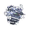 7pqlC  7pqmC  7ptfC  7ptgC 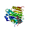 1kznS S: Starting model for refinement C: citing same article ( |
|---|---|
| Similar structure data |
-Links
 PDBj
PDBj


- Assembly
| Deposited unit | 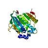
| ||||||||
|---|---|---|---|---|---|---|---|---|---|
| 1 |
| ||||||||
| Unit cell |
|
-Components
| #1: Protein | Mass: 24191.182 Da / Num. of mol.: 1 Source method: isolated from a genetically manipulated source Source: (gene. exp.) Strain: K12 Gene: gyrB, acrB, cou, himB, hisU, nalC, parA, pcbA, b3699, JW5625 Production host: References: UniProt: P0AES6, DNA topoisomerase (ATP-hydrolysing) |
|---|---|
| #2: Chemical | ChemComp-4QR / 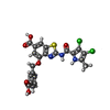  Mass: 520.342 Da / Num. of mol.: 1 / Source method: obtained synthetically / Formula: C22H15Cl2N3O6S / Feature type: SUBJECT OF INVESTIGATION Mass: 520.342 Da / Num. of mol.: 1 / Source method: obtained synthetically / Formula: C22H15Cl2N3O6S / Feature type: SUBJECT OF INVESTIGATION |
| #3: Chemical | ChemComp-TRS /   Mass: 122.143 Da / Num. of mol.: 1 / Source method: obtained synthetically / Formula: C4H12NO3 / Comment: pH buffer*YM Mass: 122.143 Da / Num. of mol.: 1 / Source method: obtained synthetically / Formula: C4H12NO3 / Comment: pH buffer*YM |
| #4: Chemical | ChemComp-MG /   Mass: 24.305 Da / Num. of mol.: 1 / Source method: obtained synthetically / Formula: Mg Mass: 24.305 Da / Num. of mol.: 1 / Source method: obtained synthetically / Formula: Mg |
| #5: Water | ChemComp-HOH /  Mass: 18.015 Da / Num. of mol.: 197 / Source method: isolated from a natural source / Formula: H2O Mass: 18.015 Da / Num. of mol.: 197 / Source method: isolated from a natural source / Formula: H2O |
| Has ligand of interest | Y |
-Experimental details
-Experiment
| Experiment | Method: X-RAY DIFFRACTION / Number of used crystals: 1 |
|---|
- Sample preparation
| Crystal | Density Matthews: 2.21 Å3/Da / Density % sol: 44.27 % |
|---|---|
| Crystal grow | Temperature: 293 K / Method: vapor diffusion, sitting drop / Details: 34% PEG4K, IOOmMTris pH8, 128mM MgC12 |
-Data collection
| Diffraction | Mean temperature: 100 K / Serial crystal experiment: N | |||||||||||||||||||||||||||
|---|---|---|---|---|---|---|---|---|---|---|---|---|---|---|---|---|---|---|---|---|---|---|---|---|---|---|---|---|
| Diffraction source | Source: SYNCHROTRON / Site: Diamond / Beamline: I03 / Wavelength: 0.9762 Å | |||||||||||||||||||||||||||
| Detector | Type: DECTRIS EIGER X 16M / Detector: PIXEL / Date: May 12, 2019 | |||||||||||||||||||||||||||
| Radiation | Protocol: SINGLE WAVELENGTH / Monochromatic (M) / Laue (L): M / Scattering type: x-ray | |||||||||||||||||||||||||||
| Radiation wavelength | Wavelength: 0.9762 Å / Relative weight: 1 | |||||||||||||||||||||||||||
| Reflection | Resolution: 1.65→52.62 Å / Num. obs: 26432 / % possible obs: 99.8 % / Redundancy: 12.9 % / Biso Wilson estimate: 18.3 Å2 / CC1/2: 0.997 / Rmerge(I) obs: 0.196 / Rpim(I) all: 0.057 / Rrim(I) all: 0.205 / Net I/σ(I): 9.4 | |||||||||||||||||||||||||||
| Reflection shell | Diffraction-ID: 1 / % possible all: 99.8
|
- Processing
| Software |
| ||||||||||||||||||||||||||||||||||||||||||||||||||||||||||||
|---|---|---|---|---|---|---|---|---|---|---|---|---|---|---|---|---|---|---|---|---|---|---|---|---|---|---|---|---|---|---|---|---|---|---|---|---|---|---|---|---|---|---|---|---|---|---|---|---|---|---|---|---|---|---|---|---|---|---|---|---|---|
| Refinement | Method to determine structure: MOLECULAR REPLACEMENT Starting model: 1kzn Resolution: 1.65→44.24 Å / Cor.coef. Fo:Fc: 0.965 / Cor.coef. Fo:Fc free: 0.947 / SU B: 5.813 / SU ML: 0.093 / Cross valid method: THROUGHOUT / σ(F): 0 / ESU R: 0.102 / ESU R Free: 0.101 / Stereochemistry target values: MAXIMUM LIKELIHOOD Details: HYDROGENS HAVE BEEN ADDED IN THE RIDING POSITIONS U VALUES : WITH TLS ADDED
| ||||||||||||||||||||||||||||||||||||||||||||||||||||||||||||
| Solvent computation | Ion probe radii: 0.8 Å / Shrinkage radii: 0.8 Å / VDW probe radii: 1.2 Å / Solvent model: MASK | ||||||||||||||||||||||||||||||||||||||||||||||||||||||||||||
| Displacement parameters | Biso max: 66.39 Å2 / Biso mean: 24.348 Å2 / Biso min: 10 Å2
| ||||||||||||||||||||||||||||||||||||||||||||||||||||||||||||
| Refinement step | Cycle: final / Resolution: 1.65→44.24 Å
| ||||||||||||||||||||||||||||||||||||||||||||||||||||||||||||
| Refine LS restraints |
| ||||||||||||||||||||||||||||||||||||||||||||||||||||||||||||
| LS refinement shell | Resolution: 1.65→1.693 Å / Rfactor Rfree error: 0 / Total num. of bins used: 20
| ||||||||||||||||||||||||||||||||||||||||||||||||||||||||||||
| Refinement TLS params. | Method: refined / Origin x: -18.857 Å / Origin y: 1.99 Å / Origin z: -4.021 Å
| ||||||||||||||||||||||||||||||||||||||||||||||||||||||||||||
| Refinement TLS group |
|
