 Movie
Movie Controller

[English] 日本語
 Yorodumi
- PDB-7omo: Crystal structure of coelenteramine-bound Renilla reniformis luci... -
+ Open data

- Basic information
| Entry | Database: PDB / ID: 7omo | ||||||
|---|---|---|---|---|---|---|---|
| Title | Crystal structure of coelenteramine-bound Renilla reniformis luciferase RLuc8-D120A variant | ||||||
 Components Components | Renilla reniformis luciferase RLuc8-D120A variant | ||||||
 Keywords Keywords | LUMINESCENT PROTEIN / bioluminscence / coelenteramide-bound enzyme | ||||||
| Function / homology | 4-[5-azanyl-6-(phenylmethyl)pyrazin-2-yl]phenol Function and homology information Function and homology information | ||||||
| Biological species |  Renilla reniformis (sea pansy) Renilla reniformis (sea pansy) | ||||||
| Method |  X-RAY DIFFRACTION / SYNCHROTRON / MOLECULAR REPLACEMENT / molecular replacement / Resolution: 1.451 Å X-RAY DIFFRACTION / SYNCHROTRON / MOLECULAR REPLACEMENT / molecular replacement / Resolution: 1.451 Å | ||||||
 Authors Authors | Schenkmayerova, A. / Marek, M. | ||||||
| Funding support |  Czech Republic, 1items Czech Republic, 1items
| ||||||
 Citation Citation | Journal: Nat Catal / Year: 2023 Title: Catalytic mechanism for Renilla-type luciferases Authors: Schenkmayerova, A. / Toul, M. / Pluskal, D. / Baatallah, R. / Gagnot, G. / Pinto, G.P. / Santana, V.T. / Stuchla, M. / Neugebauer, P. / Chaiyen, P. / Damborsky, J. / Bednar, D. / Janin, Y.L. ...Authors: Schenkmayerova, A. / Toul, M. / Pluskal, D. / Baatallah, R. / Gagnot, G. / Pinto, G.P. / Santana, V.T. / Stuchla, M. / Neugebauer, P. / Chaiyen, P. / Damborsky, J. / Bednar, D. / Janin, Y.L. / Prokop, Z. / Marek, M. | ||||||
| History |
|
- Structure visualization
| Structure viewer | Molecule: MolmilJmol/JSmol |
|---|
- Downloads & links
-Download
| PDBx/mmCIF format | 7omo.cif.gz | 147.4 KB | Display | PDBx/mmCIF format |
|---|---|---|---|---|
| PDB format | pdb7omo.ent.gz | 115 KB | Display | PDB format |
| PDBx/mmJSON format | 7omo.json.gz | Tree view | PDBx/mmJSON format | |
| Others |  Other downloads Other downloads |
-Validation report
| Summary document | 7omo_validation.pdf.gz | 2.7 MB | Display | wwPDB validaton report |
|---|---|---|---|---|
| Full document | 7omo_full_validation.pdf.gz | 2.7 MB | Display | |
| Data in XML | 7omo_validation.xml.gz | 27.1 KB | Display | |
| Data in CIF | 7omo_validation.cif.gz | 40.4 KB | Display | |
| Arichive directory | https://data.pdbj.org/pub/pdb/validation_reports/om/7omoftp://data.pdbj.org/pub/pdb/validation_reports/om/7omo | HTTPS FTP |
-Related structure data
| Related structure data | 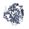 7omdC 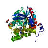 7omrC  7qxqC 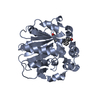 7qxrC  2spjS S: Starting model for refinement C: citing same article ( |
|---|---|
| Similar structure data |
-Links
 PDBj
PDBj- Assembly
| Deposited unit | 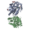
| ||||||||
|---|---|---|---|---|---|---|---|---|---|
| 1 | 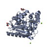
| ||||||||
| 2 | 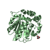
| ||||||||
| Unit cell |
|
-Components
-Protein , 1 types, 2 molecules AB
| #1: Protein | Mass: 36903.164 Da / Num. of mol.: 2 Source method: isolated from a genetically manipulated source Source: (gene. exp.) Renilla reniformis (sea pansy) / Production host:  |
|---|
-Non-polymers , 5 types, 472 molecules 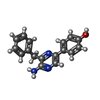


| #2: Chemical | ChemComp-VKB /  Mass: 277.321 Da / Num. of mol.: 1 / Source method: obtained synthetically / Formula: C17H15N3O / Feature type: SUBJECT OF INVESTIGATION Mass: 277.321 Da / Num. of mol.: 1 / Source method: obtained synthetically / Formula: C17H15N3O / Feature type: SUBJECT OF INVESTIGATION | ||||||
|---|---|---|---|---|---|---|---|
| #3: Chemical |  Mass: 24.305 Da / Num. of mol.: 2 / Source method: obtained synthetically / Formula: Mg / Feature type: SUBJECT OF INVESTIGATION Mass: 24.305 Da / Num. of mol.: 2 / Source method: obtained synthetically / Formula: Mg / Feature type: SUBJECT OF INVESTIGATION#4: Chemical |  Mass: 35.453 Da / Num. of mol.: 2 / Source method: obtained synthetically / Formula: Cl / Feature type: SUBJECT OF INVESTIGATION Mass: 35.453 Da / Num. of mol.: 2 / Source method: obtained synthetically / Formula: Cl / Feature type: SUBJECT OF INVESTIGATION#5: Chemical | ChemComp-GOL /  Mass: 92.094 Da / Num. of mol.: 4 / Source method: obtained synthetically / Formula: C3H8O3 / Feature type: SUBJECT OF INVESTIGATION Mass: 92.094 Da / Num. of mol.: 4 / Source method: obtained synthetically / Formula: C3H8O3 / Feature type: SUBJECT OF INVESTIGATION#6: Water | ChemComp-HOH / | Mass: 18.015 Da / Num. of mol.: 463 / Source method: isolated from a natural source / Formula: H2O |
-Details
| Has ligand of interest | Y |
|---|
-Experimental details
-Experiment
| Experiment | Method: X-RAY DIFFRACTION / Number of used crystals: 1 |
|---|
- Sample preparation
| Crystal | Density Matthews: 2 Å3/Da / Density % sol: 43.74 % |
|---|---|
| Crystal grow | Temperature: 293.15 K / Method: vapor diffusion, hanging drop / pH: 6.5 / Details: PEG3350, magnesium chloride, bis-tris |
-Data collection
| Diffraction | Mean temperature: 100 K / Serial crystal experiment: N | ||||||||||||||||||||||||
|---|---|---|---|---|---|---|---|---|---|---|---|---|---|---|---|---|---|---|---|---|---|---|---|---|---|
| Diffraction source | Source: SYNCHROTRON / Site: SLS  / Beamline: X06DA / Wavelength: 0.999 Å / Beamline: X06DA / Wavelength: 0.999 Å | ||||||||||||||||||||||||
| Detector | Type: DECTRIS PILATUS 2M-F / Detector: PIXEL / Date: Aug 27, 2018 | ||||||||||||||||||||||||
| Radiation | Protocol: SINGLE WAVELENGTH / Monochromatic (M) / Laue (L): M / Scattering type: x-ray | ||||||||||||||||||||||||
| Radiation wavelength | Wavelength: 0.999 Å / Relative weight: 1 | ||||||||||||||||||||||||
| Reflection | Resolution: 1.45→46.751 Å / Num. obs: 111521 / % possible obs: 99.9 % / Redundancy: 6.8 % / Biso Wilson estimate: 17.13 Å2 / CC1/2: 1 / Rmerge(I) obs: 0.046 / Rpim(I) all: 0.019 / Rrim(I) all: 0.049 / Net I/σ(I): 21.2 | ||||||||||||||||||||||||
| Reflection shell | Diffraction-ID: 1
|
-Phasing
| Phasing | Method: molecular replacement |
|---|
- Processing
| Software |
| |||||||||||||||||||||||||||||||||||||||||||||||||||||||||||||||||||||||||||||||||||||||||||||||||||||||||||||||||||||||||||||||||||||||||||||||||||||||||||
|---|---|---|---|---|---|---|---|---|---|---|---|---|---|---|---|---|---|---|---|---|---|---|---|---|---|---|---|---|---|---|---|---|---|---|---|---|---|---|---|---|---|---|---|---|---|---|---|---|---|---|---|---|---|---|---|---|---|---|---|---|---|---|---|---|---|---|---|---|---|---|---|---|---|---|---|---|---|---|---|---|---|---|---|---|---|---|---|---|---|---|---|---|---|---|---|---|---|---|---|---|---|---|---|---|---|---|---|---|---|---|---|---|---|---|---|---|---|---|---|---|---|---|---|---|---|---|---|---|---|---|---|---|---|---|---|---|---|---|---|---|---|---|---|---|---|---|---|---|---|---|---|---|---|---|---|---|
| Refinement | Method to determine structure: MOLECULAR REPLACEMENT Starting model: 2SPJ Resolution: 1.451→46.751 Å / SU ML: 0.13 / Cross valid method: THROUGHOUT / σ(F): 1.34 / Phase error: 18.54 / Stereochemistry target values: ML
| |||||||||||||||||||||||||||||||||||||||||||||||||||||||||||||||||||||||||||||||||||||||||||||||||||||||||||||||||||||||||||||||||||||||||||||||||||||||||||
| Solvent computation | Shrinkage radii: 0.9 Å / VDW probe radii: 1.11 Å / Solvent model: FLAT BULK SOLVENT MODEL | |||||||||||||||||||||||||||||||||||||||||||||||||||||||||||||||||||||||||||||||||||||||||||||||||||||||||||||||||||||||||||||||||||||||||||||||||||||||||||
| Displacement parameters | Biso max: 72.98 Å2 / Biso mean: 22.9199 Å2 / Biso min: 10.4 Å2 | |||||||||||||||||||||||||||||||||||||||||||||||||||||||||||||||||||||||||||||||||||||||||||||||||||||||||||||||||||||||||||||||||||||||||||||||||||||||||||
| Refinement step | Cycle: final / Resolution: 1.451→46.751 Å
| |||||||||||||||||||||||||||||||||||||||||||||||||||||||||||||||||||||||||||||||||||||||||||||||||||||||||||||||||||||||||||||||||||||||||||||||||||||||||||
| Refine LS restraints |
| |||||||||||||||||||||||||||||||||||||||||||||||||||||||||||||||||||||||||||||||||||||||||||||||||||||||||||||||||||||||||||||||||||||||||||||||||||||||||||
| LS refinement shell | Refine-ID: X-RAY DIFFRACTION / Rfactor Rfree error: 0 / % reflection obs: 100 %
|
