 Movie
Movie Controller

[English] 日本語
 Yorodumi
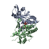
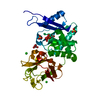
- PDB-7kvz: Structure of hSTING in complex with novel carbocyclic pyrimidine CDN-2 -
+ Open data

- Basic information
| Entry | Database: PDB / ID: 7kvz | ||||||
|---|---|---|---|---|---|---|---|
| Title | Structure of hSTING in complex with novel carbocyclic pyrimidine CDN-2 | ||||||
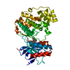
 Components Components | Stimulator of interferon genes protein | ||||||
 Keywords Keywords | IMMUNE SYSTEM / immunotherapy / stimulator of interferon genes / cyclic dinucleotide | ||||||
| Function / homology |  Function and homology information Function and homology informationautophagosome membrane / positive regulation of type I interferon production / endoplasmic reticulum-Golgi intermediate compartment membrane / activation of innate immune response / mitochondrial outer membrane / nucleotide binding / endoplasmic reticulum membrane / perinuclear region of cytoplasm Similarity search - Function | ||||||
| Biological species |  Homo sapiens (human) Homo sapiens (human) | ||||||
| Method |  X-RAY DIFFRACTION / SYNCHROTRON / MOLECULAR REPLACEMENT / Resolution: 2.35 Å X-RAY DIFFRACTION / SYNCHROTRON / MOLECULAR REPLACEMENT / Resolution: 2.35 Å | ||||||
 Authors Authors | Skene, R.J. | ||||||
 Citation Citation | Journal: J.Med.Chem. / Year: 2021 Title: Identification of Novel Carbocyclic Pyrimidine Cyclic Dinucleotide STING Agonists for Antitumor Immunotherapy Using Systemic Intravenous Route. Authors: Vyskocil, S. / Cardin, D. / Ciavarri, J. / Conlon, J. / Cullis, C. / England, D. / Gershman, R. / Gigstad, K. / Gipson, K. / Gould, A. / Greenspan, P. / Griffin, R. / Gulavita, N. / ...Authors: Vyskocil, S. / Cardin, D. / Ciavarri, J. / Conlon, J. / Cullis, C. / England, D. / Gershman, R. / Gigstad, K. / Gipson, K. / Gould, A. / Greenspan, P. / Griffin, R. / Gulavita, N. / Harrison, S. / Hu, Z. / Hu, Y. / Hata, A. / Huang, J. / Huang, S.C. / Janowick, D. / Jones, M. / Kolev, V. / Langston, S.P. / Lee, H.M. / Li, G. / Lok, D. / Ma, L. / Mai, D. / Malley, J. / Matsuda, A. / Mizutani, H. / Mizutani, M. / Molchanova, N. / Nunes, E. / Pusalkar, S. / Renou, C. / Rowland, S. / Sato, Y. / Shaw, M. / Shen, L. / Shi, Z. / Skene, R. / Soucy, F. / Stroud, S. / Xu, H. / Xu, T. / Abu-Yousif, A.O. / Zhang, J. | ||||||
| History |
|
- Structure visualization
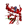
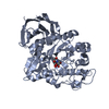
| Structure viewer | Molecule: MolmilJmol/JSmol |
|---|
- Downloads & links
-Download
| PDBx/mmCIF format | 7kvz.cif.gz | 96.1 KB | Display | PDBx/mmCIF format |
|---|---|---|---|---|
| PDB format | pdb7kvz.ent.gz | 70.6 KB | Display | PDB format |
| PDBx/mmJSON format | 7kvz.json.gz | Tree view | PDBx/mmJSON format | |
| Others |  Other downloads Other downloads |
-Validation report
| Summary document | 7kvz_validation.pdf.gz | 869.9 KB | Display | wwPDB validaton report |
|---|---|---|---|---|
| Full document | 7kvz_full_validation.pdf.gz | 870.3 KB | Display | |
| Data in XML | 7kvz_validation.xml.gz | 9.6 KB | Display | |
| Data in CIF | 7kvz_validation.cif.gz | 12.6 KB | Display | |
| Arichive directory | https://data.pdbj.org/pub/pdb/validation_reports/kv/7kvzftp://data.pdbj.org/pub/pdb/validation_reports/kv/7kvz | HTTPS FTP |
-Related structure data
| Related structure data | 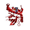 7kvxC 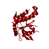 7kw1C  4ksyS S: Starting model for refinement C: citing same article ( |
|---|---|
| Similar structure data |


-Links
 PDBj
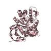
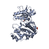
PDBj- Assembly
| Deposited unit | 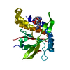
| ||||||||
|---|---|---|---|---|---|---|---|---|---|
| 1 | 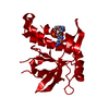
| ||||||||
| Unit cell |
|
-Components
| #1: Protein | Mass: 27211.619 Da / Num. of mol.: 1 Source method: isolated from a genetically manipulated source Source: (gene. exp.) Homo sapiens (human) / Gene: STING, LOC340061, hCG_1782396 / Production host:  |
|---|---|
| #2: Chemical | ChemComp-X5D / (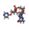  Mass: 617.400 Da / Num. of mol.: 1 / Source method: obtained synthetically / Formula: C20H25N7O12P2 / Feature type: SUBJECT OF INVESTIGATION Mass: 617.400 Da / Num. of mol.: 1 / Source method: obtained synthetically / Formula: C20H25N7O12P2 / Feature type: SUBJECT OF INVESTIGATION |
| #3: Water | ChemComp-HOH /  Mass: 18.015 Da / Num. of mol.: 60 / Source method: isolated from a natural source / Formula: H2O Mass: 18.015 Da / Num. of mol.: 60 / Source method: isolated from a natural source / Formula: H2O |
| Has ligand of interest | Y |
-Experimental details
-Experiment
| Experiment | Method: X-RAY DIFFRACTION / Number of used crystals: 1 |
|---|
- Sample preparation
| Crystal | Density Matthews: 2.3 Å3/Da / Density % sol: 46.41 % |
|---|---|
| Crystal grow | Temperature: 293 K / Method: vapor diffusion, sitting drop / Details: 20.0% PEG 3350, 0.2M sodium formate |
-Data collection
| Diffraction | Mean temperature: 100 K / Serial crystal experiment: N | ||||||||||||||||||||||||||||||
|---|---|---|---|---|---|---|---|---|---|---|---|---|---|---|---|---|---|---|---|---|---|---|---|---|---|---|---|---|---|---|---|
| Diffraction source | Source: SYNCHROTRON / Site: ALS  / Beamline: 5.0.2 / Wavelength: 0.987 Å / Beamline: 5.0.2 / Wavelength: 0.987 Å | ||||||||||||||||||||||||||||||
| Detector | Type: DECTRIS PILATUS3 S 6M / Detector: PIXEL / Date: Aug 1, 2017 | ||||||||||||||||||||||||||||||
| Radiation | Protocol: SINGLE WAVELENGTH / Monochromatic (M) / Laue (L): M / Scattering type: x-ray | ||||||||||||||||||||||||||||||
| Radiation wavelength | Wavelength: 0.987 Å / Relative weight: 1 | ||||||||||||||||||||||||||||||
| Reflection | Resolution: 2.35→44.45 Å / Num. obs: 9973 / % possible obs: 96.6 % / Redundancy: 3.3 % / CC1/2: 0.997 / Rmerge(I) obs: 0.072 / Rpim(I) all: 0.047 / Rrim(I) all: 0.086 / Net I/σ(I): 8.2 | ||||||||||||||||||||||||||||||
| Reflection shell | Diffraction-ID: 1
|
- Processing
| Software |
| |||||||||||||||||||||||||||||||||||||||||||||
|---|---|---|---|---|---|---|---|---|---|---|---|---|---|---|---|---|---|---|---|---|---|---|---|---|---|---|---|---|---|---|---|---|---|---|---|---|---|---|---|---|---|---|---|---|---|---|
| Refinement | Method to determine structure: MOLECULAR REPLACEMENT Starting model: 4KSY Resolution: 2.35→44.446 Å / Cor.coef. Fo:Fc: 0.966 / Cor.coef. Fo:Fc free: 0.938 / SU B: 16.783 / SU ML: 0.172 / SU R Cruickshank DPI: 0.2948 / Cross valid method: THROUGHOUT / σ(F): 0 / ESU R: 0.295 / ESU R Free: 0.226 / Stereochemistry target values: MAXIMUM LIKELIHOOD / Details: U VALUES : WITH TLS ADDED
| |||||||||||||||||||||||||||||||||||||||||||||
| Solvent computation | Ion probe radii: 0.8 Å / Shrinkage radii: 0.8 Å / VDW probe radii: 1.2 Å / Solvent model: MASK | |||||||||||||||||||||||||||||||||||||||||||||
| Displacement parameters | Biso max: 134.41 Å2 / Biso mean: 59.663 Å2 / Biso min: 19.78 Å2
| |||||||||||||||||||||||||||||||||||||||||||||
| Refinement step | Cycle: final / Resolution: 2.35→44.446 Å
| |||||||||||||||||||||||||||||||||||||||||||||
| Refine LS restraints |
| |||||||||||||||||||||||||||||||||||||||||||||
| LS refinement shell | Resolution: 2.35→2.411 Å / Rfactor Rfree error: 0 / Total num. of bins used: 20
| |||||||||||||||||||||||||||||||||||||||||||||
| Refinement TLS params. | Method: refined / Origin x: 29.789 Å / Origin y: 7.958 Å / Origin z: 17.327 Å
| |||||||||||||||||||||||||||||||||||||||||||||
| Refinement TLS group |
|
