Movie
Movie Controller

[English] 日本語
 Yorodumi
- PDB-7eoq: Structure of the human GluN1/GluN2A NMDA receptor in the glycine/... -
+ Open data

- Basic information
| Entry | Database: PDB / ID: 7eoq | ||||||||||||||||||
|---|---|---|---|---|---|---|---|---|---|---|---|---|---|---|---|---|---|---|---|
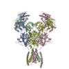
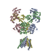
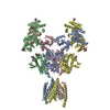
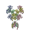
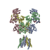
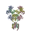
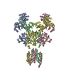
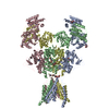
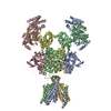
| Title | Structure of the human GluN1/GluN2A NMDA receptor in the glycine/CPP bound state | ||||||||||||||||||
 Components Components |
| ||||||||||||||||||
 Keywords Keywords | MEMBRANE PROTEIN / NMDA receptor | ||||||||||||||||||
| Function / homology |  Function and homology information Function and homology informationglycine-gated cation channel activity / directional locomotion / excitatory chemical synaptic transmission / Synaptic adhesion-like molecules / serotonin metabolic process / protein localization to postsynaptic membrane / sleep / propylene metabolic process / response to glycine / Assembly and cell surface presentation of NMDA receptors ...glycine-gated cation channel activity / directional locomotion / excitatory chemical synaptic transmission / Synaptic adhesion-like molecules / serotonin metabolic process / protein localization to postsynaptic membrane / sleep / propylene metabolic process / response to glycine / Assembly and cell surface presentation of NMDA receptors / neurotransmitter receptor complex / Neurexins and neuroligins / regulation of monoatomic cation transmembrane transport / NMDA glutamate receptor activity / NMDA selective glutamate receptor complex / glutamate binding / ligand-gated sodium channel activity / glutamate receptor signaling pathway / calcium ion transmembrane import into cytosol / protein heterotetramerization / positive regulation of reactive oxygen species biosynthetic process / glycine binding / dopamine metabolic process / startle response / Negative regulation of NMDA receptor-mediated neuronal transmission / Unblocking of NMDA receptors, glutamate binding and activation / regulation of neuronal synaptic plasticity / Long-term potentiation / positive regulation of calcium ion transport into cytosol / monoatomic cation transmembrane transport / monoatomic cation transport / ligand-gated monoatomic ion channel activity / calcium ion homeostasis / synaptic cleft / MECP2 regulates neuronal receptors and channels / positive regulation of synaptic transmission, glutamatergic / EPHB-mediated forward signaling / glutamate-gated calcium ion channel activity / neurogenesis / ionotropic glutamate receptor signaling pathway / excitatory synapse / sensory perception of pain / cytoplasmic vesicle membrane / Ras activation upon Ca2+ influx through NMDA receptor / response to amphetamine / excitatory postsynaptic potential / protein catabolic process / positive regulation of excitatory postsynaptic potential / sodium ion transmembrane transport / synaptic membrane / synaptic transmission, glutamatergic / regulation of membrane potential / transmitter-gated monoatomic ion channel activity involved in regulation of postsynaptic membrane potential / brain development / visual learning / negative regulation of protein catabolic process / regulation of synaptic plasticity / response to wounding / postsynaptic density membrane / memory / long-term synaptic potentiation / calcium ion transmembrane transport / terminal bouton / synaptic vesicle / amyloid-beta binding / signaling receptor activity / RAF/MAP kinase cascade / presynaptic membrane / chemical synaptic transmission / dendritic spine / response to ethanol / learning or memory / calmodulin binding / postsynaptic membrane / neuron projection / postsynaptic density / response to xenobiotic stimulus / positive regulation of apoptotic process / calcium ion binding / dendrite / synapse / endoplasmic reticulum membrane / protein-containing complex binding / glutamatergic synapse / cell surface / positive regulation of transcription by RNA polymerase II / zinc ion binding / plasma membrane / cytoplasm Similarity search - Function | ||||||||||||||||||
| Biological species |  Homo sapiens (human) Homo sapiens (human) | ||||||||||||||||||
| Method | ELECTRON MICROSCOPY / single particle reconstruction / cryo EM / Resolution: 4.1 Å | ||||||||||||||||||
 Authors Authors | Wang, H. / Zhu, S. | ||||||||||||||||||
| Funding support |  China, European Union, 5items China, European Union, 5items
| ||||||||||||||||||
 Citation Citation | Journal: Neuron / Year: 2021 Title: Gating mechanism and a modulatory niche of human GluN1-GluN2A NMDA receptors. Authors: Han Wang / Shiyun Lv / David Stroebel / Jinbao Zhang / Yijie Pan / Xuejing Huang / Xing Zhang / Pierre Paoletti / Shujia Zhu /  Abstract: N-methyl-D-aspartate (NMDA) receptors are glutamate-gated calcium-permeable ion channels that are widely implicated in synaptic transmission and plasticity. Here, we report a gallery of cryo-electron ...N-methyl-D-aspartate (NMDA) receptors are glutamate-gated calcium-permeable ion channels that are widely implicated in synaptic transmission and plasticity. Here, we report a gallery of cryo-electron microscopy (cryo-EM) structures of the human GluN1-GluN2A NMDA receptor at an overall resolution of 4 Å in complex with distinct ligands or modulators. In the full-length context of GluN1-GluN2A receptors, we visualize the competitive antagonists bound to the ligand-binding domains (LBDs) of GluN1 and GluN2A subunits, respectively. We reveal that the binding of positive allosteric modulator shortens the distance between LBDs and the transmembrane domain (TMD), which further stretches the opening of the gate. In addition, we unexpectedly visualize the binding cavity of the "foot-in-the-door" blocker 9-aminoacridine within the LBD-TMD linker region, differing from the conventional "trapping" blocker binding site at the vestibule within the TMD. Our study provides molecular insights into the crosstalk between LBDs and TMD during channel activation, inhibition, and allosteric transition. | ||||||||||||||||||
| History |
|
- Structure visualization
| Movie |
Movie viewer |
|---|---|
| Structure viewer | Molecule: MolmilJmol/JSmol |
- Downloads & links
-Download
| PDBx/mmCIF format | 7eoq.cif.gz | 550.2 KB | Display | PDBx/mmCIF format |
|---|---|---|---|---|
| PDB format | pdb7eoq.ent.gz | 448.2 KB | Display | PDB format |
| PDBx/mmJSON format | 7eoq.json.gz | Tree view | PDBx/mmJSON format | |
| Others |  Other downloads Other downloads |
-Validation report
| Arichive directory | https://data.pdbj.org/pub/pdb/validation_reports/eo/7eoqftp://data.pdbj.org/pub/pdb/validation_reports/eo/7eoq | HTTPS FTP |
|---|
-Related structure data
| Related structure data |  31227MC  7eorC  7eosC  7eotC  7eouC M: map data used to model this data C: citing same article ( |
|---|---|
| Similar structure data |


-Links
 PDBj
PDBj


- Assembly
| Deposited unit | 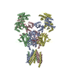
|
|---|---|
| 1 |
|
-Components
| #1: Protein | Mass: 95537.703 Da / Num. of mol.: 2 / Mutation: L794C Source method: isolated from a genetically manipulated source Source: (gene. exp.) Homo sapiens (human) / Gene: GRIN2A, NMDAR2A / Cell line (production host): HEK293S / Production host: Homo sapiens (human) / References: UniProt: Q12879#2: Protein | Mass: 95210.102 Da / Num. of mol.: 2 / Mutation: E698C Source method: isolated from a genetically manipulated source Source: (gene. exp.) Homo sapiens (human) / Gene: GRIN1, NMDAR1 / Cell line (production host): HEK293S / Production host: Homo sapiens (human) / References: UniProt: Q05586#3: Sugar | ChemComp-NAG /   Type: D-saccharide, beta linking / Mass: 221.208 Da / Num. of mol.: 8 / Source method: obtained synthetically / Formula: C8H15NO6 / Feature type: SUBJECT OF INVESTIGATION Type: D-saccharide, beta linking / Mass: 221.208 Da / Num. of mol.: 8 / Source method: obtained synthetically / Formula: C8H15NO6 / Feature type: SUBJECT OF INVESTIGATION#4: Chemical |   Mass: 252.205 Da / Num. of mol.: 2 / Source method: obtained synthetically / Formula: C8H17N2O5P / Feature type: SUBJECT OF INVESTIGATION Mass: 252.205 Da / Num. of mol.: 2 / Source method: obtained synthetically / Formula: C8H17N2O5P / Feature type: SUBJECT OF INVESTIGATIONHas ligand of interest | Y | Has protein modification | Y | |
|---|
-Experimental details
-Experiment
| Experiment | Method: ELECTRON MICROSCOPY |
|---|---|
| EM experiment | Aggregation state: PARTICLE / 3D reconstruction method: single particle reconstruction |
- Sample preparation
| Component | Name: Structure of the human GluN1/GluN2A NMDA receptor in the glycine/CPP bound state Type: COMPLEX / Entity ID: #1-#2 / Source: RECOMBINANT |
|---|---|
| Source (natural) | Organism: Homo sapiens (human) |
| Source (recombinant) | Organism: Homo sapiens (human) / Cell: HEK293S |
| Buffer solution | pH: 8 |
| Specimen | Embedding applied: NO / Shadowing applied: NO / Staining applied: NO / Vitrification applied: YES |
| Specimen support | Grid material: GOLD / Grid mesh size: 200 divisions/in. / Grid type: Quantifoil R1.2/1.3 |
| Vitrification | Instrument: FEI VITROBOT MARK III / Cryogen name: ETHANE / Humidity: 100 % / Chamber temperature: 281 K |
- Electron microscopy imaging
| Experimental equipment |  Model: Titan Krios / Image courtesy: FEI Company |
|---|---|
| Microscopy | Model: FEI TITAN KRIOS |
| Electron gun | Electron source:  FIELD EMISSION GUN / Accelerating voltage: 300 kV / Illumination mode: SPOT SCAN FIELD EMISSION GUN / Accelerating voltage: 300 kV / Illumination mode: SPOT SCAN |
| Electron lens | Mode: BRIGHT FIELD |
| Image recording | Electron dose: 60 e/Å2 / Detector mode: SUPER-RESOLUTION / Film or detector model: GATAN K2 SUMMIT (4k x 4k) |
- Processing
| Software | Name: PHENIX / Version: 1.19_4092: / Classification: refinement | ||||||||||||||||||||||||||||||||||||
|---|---|---|---|---|---|---|---|---|---|---|---|---|---|---|---|---|---|---|---|---|---|---|---|---|---|---|---|---|---|---|---|---|---|---|---|---|---|
| EM software |
| ||||||||||||||||||||||||||||||||||||
| CTF correction | Type: PHASE FLIPPING ONLY | ||||||||||||||||||||||||||||||||||||
| Symmetry | Point symmetry: C2 (2 fold cyclic) | ||||||||||||||||||||||||||||||||||||
| 3D reconstruction | Resolution: 4.1 Å / Resolution method: FSC 0.143 CUT-OFF / Num. of particles: 63459 / Symmetry type: POINT | ||||||||||||||||||||||||||||||||||||
| Atomic model building | Protocol: RIGID BODY FIT / Space: REAL | ||||||||||||||||||||||||||||||||||||
| Atomic model building | PDB-ID: 6IRA Accession code: 6IRA / Source name: PDB / Type: experimental model | ||||||||||||||||||||||||||||||||||||
| Refine LS restraints |
|