 ムービー
ムービー コントローラー


+ データを開く

- 基本情報
基本情報
| 登録情報 | データベース: PDB / ID: 6sue | ||||||
|---|---|---|---|---|---|---|---|
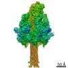
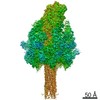
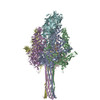
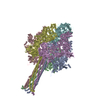
| タイトル | Structure of Photorhabdus luminescens Tc holotoxin pore, Mutation TccC3-D651A | ||||||
 要素 要素 |
| ||||||
 キーワード キーワード | TOXIN / Complex / Holotoxin / Photorhabdus / Insecticidal | ||||||
| 機能・相同性 |  機能・相同性情報 機能・相同性情報 | ||||||
| 生物種 |  Photorhabdus luminescens (バクテリア) Photorhabdus luminescens (バクテリア) | ||||||
| 手法 | 電子顕微鏡法 / 単粒子再構成法 / クライオ電子顕微鏡法 / 解像度: 3.4 Å | ||||||
 データ登録者 データ登録者 | Roderer, D. / Raunser, S. | ||||||
| 資金援助 |  ドイツ, 1件 ドイツ, 1件
| ||||||
 引用 引用 | ジャーナル: Proc Natl Acad Sci U S A / 年: 2019 タイトル: Structure of a Tc holotoxin pore provides insights into the translocation mechanism. 著者: Daniel Roderer / Oliver Hofnagel / Roland Benz / Stefan Raunser / 要旨: Tc toxins are modular toxin systems of insect and human pathogenic bacteria. They are composed of a 1.4-MDa pentameric membrane translocator (TcA) and a 250-kDa cocoon (TcB and TcC) encapsulating the ...Tc toxins are modular toxin systems of insect and human pathogenic bacteria. They are composed of a 1.4-MDa pentameric membrane translocator (TcA) and a 250-kDa cocoon (TcB and TcC) encapsulating the 30-kDa toxic enzyme (C terminus of TcC). Binding of Tc toxins to target cells and a pH shift trigger the conformational transition from the soluble prepore state to the membrane-embedded pore. Subsequently, the toxic enzyme is translocated and released into the cytoplasm. A high-resolution structure of a holotoxin embedded in membranes is missing, leaving open the question of whether TcB-TcC has an influence on the conformational transition of TcA. Here we show in atomic detail a fully assembled 1.7-MDa Tc holotoxin complex from in the membrane. We find that the 5 TcA protomers conformationally adapt to fit around the cocoon during the prepore-to-pore transition. The architecture of the Tc toxin complex allows TcB-TcC to bind to an already membrane-embedded TcA pore to form a holotoxin. Importantly, assembly of the holotoxin at the membrane results in spontaneous translocation of the toxic enzyme, indicating that this process is not driven by a proton gradient or other energy source. Mammalian lipids with zwitterionic head groups are preferred over other lipids for the integration of Tc toxins. In a nontoxic Tc toxin variant, we can visualize part of the translocating toxic enzyme, which transiently interacts with alternating negative charges and hydrophobic stretches of the translocation channel, providing insights into the mechanism of action of Tc toxins. #1: ジャーナル: Biorxivタイトル: Structure of a Tc holotoxin pore provides insights into the translocation mechanism 著者: Roderer, D. / Hofnagel, O. / Benz, R. / Raunser, S. | ||||||
| 履歴 |
|
- 構造の表示
構造の表示
| ムービー |
ムービービューア |
|---|---|
| 構造ビューア | 分子: MolmilJmol/JSmol |
- ダウンロードとリンク
-ダウンロード
| PDBx/mmCIF形式 | 6sue.cif.gz | 2.3 MB | 表示 | PDBx/mmCIF形式 |
|---|---|---|---|---|
| PDB形式 | pdb6sue.ent.gz | 表示 | PDB形式 | |
| PDBx/mmJSON形式 | 6sue.json.gz | ツリー表示 | PDBx/mmJSON形式 | |
| その他 |  その他のダウンロード その他のダウンロード |
-検証レポート
| アーカイブディレクトリ | https://data.pdbj.org/pub/pdb/validation_reports/su/6sueftp://data.pdbj.org/pub/pdb/validation_reports/su/6sue | HTTPS FTP |
|---|
-関連構造データ

-リンク
 PDBj
PDBj
- 集合体
集合体
| 登録構造単位 | 
|
|---|---|
| 1 |
|
-要素
| #1: タンパク質 | 分子量: 283230.375 Da / 分子数: 5 / 由来タイプ: 組換発現 由来: (組換発現) Photorhabdus luminescens (バクテリア)遺伝子: tcdA, tcdA1 / 発現宿主: #2: タンパク質 | | 分子量: 274144.250 Da / 分子数: 1 / 由来タイプ: 組換発現 由来: (組換発現) Photorhabdus luminescens (バクテリア)遺伝子: tcdB2, TccC3 / 発現宿主: |
|---|
-実験情報
-実験
| 実験 | 手法: 電子顕微鏡法 |
|---|---|
| EM実験 | 試料の集合状態: PARTICLE / 3次元再構成法: 単粒子再構成法 |
- 試料調製
試料調製
| 構成要素 | 名称: Tc holotoxin composed of the TcdA1 pentamer and TcdB2-TccC3 タイプ: COMPLEX / 詳細: Mutation D651A in TccC3 / Entity ID: all / 由来: RECOMBINANT |
|---|---|
| 分子量 | 値: 1.7 MDa / 実験値: NO |
| 由来(天然) | 生物種: Photorhabdus luminescens (バクテリア) |
| 由来(組換発現) | 生物種: |
| 緩衝液 | pH: 8 |
| 試料 | 濃度: 0.1 mg/ml / 包埋: NO / シャドウイング: NO / 染色: NO / 凍結: YES |
| 急速凍結 | 凍結剤: ETHANE |
- 電子顕微鏡撮影
| 実験機器 |  モデル: Titan Krios / 画像提供: FEI Company |
|---|---|
| 顕微鏡 | モデル: FEI TITAN KRIOS |
| 電子銃 | 電子線源:  FIELD EMISSION GUN / 加速電圧: 300 kV / 照射モード: SPOT SCAN FIELD EMISSION GUN / 加速電圧: 300 kV / 照射モード: SPOT SCAN |
| 電子レンズ | モード: BRIGHT FIELD |
| 撮影 | 電子線照射量: 100 e/Å2 / 検出モード: INTEGRATING フィルム・検出器のモデル: FEI FALCON III (4k x 4k) |
- 解析
解析
| EMソフトウェア | 名称: SPHIRE / カテゴリ: 3次元再構成 |
|---|---|
| CTF補正 | タイプ: PHASE FLIPPING AND AMPLITUDE CORRECTION |
| 対称性 | 点対称性: C1 (非対称) |
| 3次元再構成 | 解像度: 3.4 Å / 解像度の算出法: FSC 0.143 CUT-OFF / 粒子像の数: 337823 / 対称性のタイプ: POINT |
