 Movie
Movie Controller

[English] 日本語
 Yorodumi
- PDB-6r83: CryoEM structure and molecular model of squid hemocyanin (Todarod... -
+ Open data

- Basic information
| Entry | Database: PDB / ID: 6r83 | ||||||||||||
|---|---|---|---|---|---|---|---|---|---|---|---|---|---|
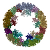
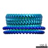
| Title | CryoEM structure and molecular model of squid hemocyanin (Todarodes pacificus , TpH) | ||||||||||||
 Components Components | Hemocyanin subunit 1 | ||||||||||||
 Keywords Keywords | OXYGEN TRANSPORT / oxygen transporter / mollusc / hemocyanin / copper / symmetry mismatch | ||||||||||||
| Function / homology |  Function and homology information Function and homology information | ||||||||||||
| Biological species |  Todarodes pacificus (Japanese flying squid) Todarodes pacificus (Japanese flying squid) | ||||||||||||
| Method | ELECTRON MICROSCOPY / single particle reconstruction / cryo EM / Resolution: 5.1 Å | ||||||||||||
 Authors Authors | Tanaka, Y. / Kato, S. / Stabrin, M. / Raunser, S. / Matsui, T. / Gatsogiannis, C. | ||||||||||||
| Funding support |  Germany, Germany,  Japan, 3items Japan, 3items
| ||||||||||||
 Citation Citation | Journal: IUCrJ / Year: 2019 Title: Cryo-EM reveals the asymmetric assembly of squid hemocyanin. Authors: Yoshikazu Tanaka / Sanae Kato / Markus Stabrin / Stefan Raunser / Takashi Matsui / Christos Gatsogiannis / Abstract: The oxygen transporter of molluscs, hemocyanin, consists of long pearl-necklace-like subunits of several globular domains. The subunits assemble in a complex manner to form cylindrical decamers. ...The oxygen transporter of molluscs, hemocyanin, consists of long pearl-necklace-like subunits of several globular domains. The subunits assemble in a complex manner to form cylindrical decamers. Typically, the first six domains of each subunit assemble together to form the cylinder wall, while the C-terminal domains form a collar that fills or caps the cylinder. During evolution, various molluscs have been able to fine-tune their oxygen binding by deleting or adding C-terminal domains and adjusting their inner-collar architecture. However, squids have duplicated one of the wall domains of their subunits instead. Here, using cryo-EM and an optimized refinement protocol implemented in , this work tackled the symmetry-mismatched structure of squid hemocyanin, revealing the precise effect of this duplication on its quaternary structure and providing a potential model for its structural evolution. | ||||||||||||
| History |
|
- Structure visualization
| Movie |
Movie viewer |
|---|---|
| Structure viewer | Molecule: MolmilJmol/JSmol |
- Downloads & links
-Download
| PDBx/mmCIF format | 6r83.cif.gz | 4.8 MB | Display | PDBx/mmCIF format |
|---|---|---|---|---|
| PDB format | pdb6r83.ent.gz | Display | PDB format | |
| PDBx/mmJSON format | 6r83.json.gz | Tree view | PDBx/mmJSON format | |
| Others |  Other downloads Other downloads |
-Validation report
| Arichive directory | https://data.pdbj.org/pub/pdb/validation_reports/r8/6r83ftp://data.pdbj.org/pub/pdb/validation_reports/r8/6r83 | HTTPS FTP |
|---|
-Related structure data
| Related structure data | 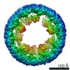 4750MC M: map data used to model this data C: citing same article ( |
|---|---|
| Similar structure data |
-Links
 PDBj
PDBj- Assembly
| Deposited unit | 
|
|---|---|
| 1 |
|
-Components
| #1: Protein | Mass: 379741.188 Da / Num. of mol.: 10 / Source method: isolated from a natural source Source: (natural) Todarodes pacificus (Japanese flying squid)References: UniProt: A0A0P0UX03 |
|---|
-Experimental details
-Experiment
| Experiment | Method: ELECTRON MICROSCOPY |
|---|---|
| EM experiment | Aggregation state: PARTICLE / 3D reconstruction method: single particle reconstruction |
- Sample preparation
| Component | Name: Squid Hemocyanin / Type: COMPLEX Details: TpH is a decamer of a 400 kDa subunit. Each subunit is composed by 8 paralogous O2 binding functional units (A,B,C,D,D*,E,F,G). The ten subunits of TpH acquire 4 different overall conformations. Entity ID: all / Source: NATURAL | |||||||||||||||
|---|---|---|---|---|---|---|---|---|---|---|---|---|---|---|---|---|
| Molecular weight | Value: 3.8 MDa / Experimental value: NO | |||||||||||||||
| Source (natural) | Organism: Todarodes pacificus (Japanese flying squid) | |||||||||||||||
| Buffer solution | pH: 7.5 | |||||||||||||||
| Buffer component |
| |||||||||||||||
| Specimen | Conc.: 2.9 mg/ml / Embedding applied: NO / Shadowing applied: NO / Staining applied: NO / Vitrification applied: YES | |||||||||||||||
| Specimen support | Grid material: COPPER / Grid type: Quantifoil R2/1 | |||||||||||||||
| Vitrification | Instrument: GATAN CRYOPLUNGE 3 / Cryogen name: ETHANE |
- Electron microscopy imaging
| Experimental equipment |  Model: Titan Krios / Image courtesy: FEI Company |
|---|---|
| Microscopy | Model: FEI TITAN KRIOS |
| Electron gun | Electron source:  FIELD EMISSION GUN / Accelerating voltage: 300 kV / Illumination mode: OTHER FIELD EMISSION GUN / Accelerating voltage: 300 kV / Illumination mode: OTHER |
| Electron lens | Mode: BRIGHT FIELD / Cs: 0 mm |
| Specimen holder | Cryogen: NITROGEN / Specimen holder model: FEI TITAN KRIOS AUTOGRID HOLDER |
| Image recording | Average exposure time: 1 sec. / Electron dose: 56 e/Å2 / Film or detector model: FEI FALCON II (4k x 4k) / Num. of grids imaged: 1 / Num. of real images: 5092 |
| EM imaging optics | Spherical aberration corrector: Microscope equipped with Cs corrector |
| Image scans | Movie frames/image: 24 / Used frames/image: 1-24 |
- Processing
| EM software |
| ||||||||||||||||||||||||||||
|---|---|---|---|---|---|---|---|---|---|---|---|---|---|---|---|---|---|---|---|---|---|---|---|---|---|---|---|---|---|
| CTF correction | Type: PHASE FLIPPING AND AMPLITUDE CORRECTION | ||||||||||||||||||||||||||||
| Particle selection | Num. of particles selected: 359250 | ||||||||||||||||||||||||||||
| Symmetry | Point symmetry: C1 (asymmetric) | ||||||||||||||||||||||||||||
| 3D reconstruction | Resolution: 5.1 Å / Resolution method: FSC 0.143 CUT-OFF / Num. of particles: 196315 / Symmetry type: POINT | ||||||||||||||||||||||||||||
| Atomic model building | Protocol: RIGID BODY FIT / Target criteria: Cross-correlation coefficient |
