 Movie
Movie Controller


+ Open data

- Basic information
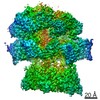
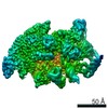
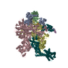
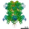
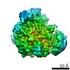
| Entry | Database: PDB / ID: 6qem | ||||||||||||
|---|---|---|---|---|---|---|---|---|---|---|---|---|---|
| Title | E. coli DnaBC complex bound to ssDNA | ||||||||||||
 Components Components |
| ||||||||||||
 Keywords Keywords | REPLICATION / Helicase / helicase loader / AAA+ / RecA | ||||||||||||
| Function / homology |  Function and homology information Function and homology informationDnaB-DnaC complex / DnaB-DnaC-Rep-PriC complex / DnaB-DnaG complex / DnaB-DnaC-DnaT-PriA-PriC complex / DnaB-DnaC-DnaT-PriA-PriB complex / DNA helicase complex / primosome complex / DNA 5'-3' helicase / DNA replication, synthesis of primer / replisome ...DnaB-DnaC complex / DnaB-DnaC-Rep-PriC complex / DnaB-DnaG complex / DnaB-DnaC-DnaT-PriA-PriC complex / DnaB-DnaC-DnaT-PriA-PriB complex / DNA helicase complex / primosome complex / DNA 5'-3' helicase / DNA replication, synthesis of primer / replisome / response to ionizing radiation / DNA strand elongation involved in DNA replication / replication fork processing / DNA replication initiation / DNA helicase activity / helicase activity / Hydrolases; Acting on acid anhydrides; Acting on acid anhydrides to facilitate cellular and subcellular movement / 5'-3' DNA helicase activity / DNA replication / ATP hydrolysis activity / DNA binding / ATP binding / identical protein binding / cytosol Similarity search - Function | ||||||||||||
| Biological species |  | ||||||||||||
| Method | ELECTRON MICROSCOPY / single particle reconstruction / cryo EM / Resolution: 3.4 Å | ||||||||||||
 Authors Authors | Arias-Palomo, E. / Puri, N. / O'Shea Murray, V.L. / Yan, Q. / Berger, J.M. | ||||||||||||
| Funding support |  United States, United States,  Spain, 3items Spain, 3items
| ||||||||||||
 Citation Citation | Journal: Mol Cell / Year: 2019 Title: Physical Basis for the Loading of a Bacterial Replicative Helicase onto DNA. Authors: Ernesto Arias-Palomo / Neha Puri / Valerie L O'Shea Murray / Qianyun Yan / James M Berger / Abstract: In cells, dedicated AAA+ ATPases deposit hexameric, ring-shaped helicases onto DNA to initiate chromosomal replication. To better understand the mechanisms by which helicase loading can occur, we ...In cells, dedicated AAA+ ATPases deposit hexameric, ring-shaped helicases onto DNA to initiate chromosomal replication. To better understand the mechanisms by which helicase loading can occur, we used cryo-EM to determine sub-4-Å-resolution structures of the E. coli DnaB⋅DnaC helicase⋅loader complex with nucleotide in pre- and post-DNA engagement states. In the absence of DNA, six DnaC protomers latch onto and crack open a DnaB hexamer using an extended N-terminal domain, stabilizing this conformation through nucleotide-dependent ATPase interactions. Upon binding DNA, DnaC hydrolyzes ATP, allowing DnaB to isomerize into a topologically closed, pre-translocation state competent to bind primase. Our data show how DnaC opens the DnaB ring and represses the helicase prior to DNA binding and how DnaC ATPase activity is reciprocally regulated by DnaB and DNA. Comparative analyses reveal how the helicase loading mechanism of DnaC parallels and diverges from homologous AAA+ systems involved in DNA replication and transposition. | ||||||||||||
| History |
|
- Structure visualization
| Movie |
Movie viewer |
|---|---|
| Structure viewer | Molecule: MolmilJmol/JSmol |
- Downloads & links
-Download
| PDBx/mmCIF format | 6qem.cif.gz | 1.3 MB | Display | PDBx/mmCIF format |
|---|---|---|---|---|
| PDB format | pdb6qem.ent.gz | 1.1 MB | Display | PDB format |
| PDBx/mmJSON format | 6qem.json.gz | Tree view | PDBx/mmJSON format | |
| Others |  Other downloads Other downloads |
-Validation report
| Summary document | 6qem_validation.pdf.gz | 1.6 MB | Display | wwPDB validaton report |
|---|---|---|---|---|
| Full document | 6qem_full_validation.pdf.gz | 1.6 MB | Display | |
| Data in XML | 6qem_validation.xml.gz | 114.4 KB | Display | |
| Data in CIF | 6qem_validation.cif.gz | 168.3 KB | Display | |
| Arichive directory | https://data.pdbj.org/pub/pdb/validation_reports/qe/6qemftp://data.pdbj.org/pub/pdb/validation_reports/qe/6qem | HTTPS FTP |
-Related structure data
| Related structure data |  4538MC  4537C  6qelC C: citing same article ( M: map data used to model this data |
|---|---|
| Similar structure data |

-Links
 PDBj
PDBj


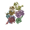
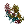
- Assembly
| Deposited unit | 
|
|---|---|
| 1 |
|
-Components
-Protein , 2 types, 12 molecules ABCDEFGHIJKL
| #1: Protein | Mass: 52450.945 Da / Num. of mol.: 6 Source method: isolated from a genetically manipulated source Source: (gene. exp.) #2: Protein | Mass: 27973.152 Da / Num. of mol.: 6 Source method: isolated from a genetically manipulated source Source: (gene. exp.) |
|---|
-DNA chain , 1 types, 1 molecules M
| #3: DNA chain | Mass: 10905.983 Da / Num. of mol.: 1 / Source method: obtained synthetically / Source: (synth.) |
|---|
-Non-polymers , 3 types, 16 molecules 


| #4: Chemical | ChemComp-08T / [[[(  Mass: 492.201 Da / Num. of mol.: 5 / Source method: obtained synthetically / Formula: C10H14BeF3N5O10P2 Mass: 492.201 Da / Num. of mol.: 5 / Source method: obtained synthetically / Formula: C10H14BeF3N5O10P2#5: Chemical | ChemComp-MG /  Mass: 24.305 Da / Num. of mol.: 5 / Source method: obtained synthetically / Formula: Mg Mass: 24.305 Da / Num. of mol.: 5 / Source method: obtained synthetically / Formula: Mg#6: Chemical | ChemComp-ADP /  Mass: 427.201 Da / Num. of mol.: 6 / Source method: obtained synthetically / Formula: C10H15N5O10P2 / Comment: ADP, energy-carrying molecule*YM Mass: 427.201 Da / Num. of mol.: 6 / Source method: obtained synthetically / Formula: C10H15N5O10P2 / Comment: ADP, energy-carrying molecule*YM |
|---|
-Experimental details
-Experiment
| Experiment | Method: ELECTRON MICROSCOPY |
|---|---|
| EM experiment | Aggregation state: PARTICLE / 3D reconstruction method: single particle reconstruction |
- Sample preparation
| Component |
| ||||||||||||||||||||||||
|---|---|---|---|---|---|---|---|---|---|---|---|---|---|---|---|---|---|---|---|---|---|---|---|---|---|
| Molecular weight | Value: 0.49 MDa / Experimental value: NO | ||||||||||||||||||||||||
| Source (natural) |
| ||||||||||||||||||||||||
| Source (recombinant) |
| ||||||||||||||||||||||||
| Buffer solution | pH: 7.5 | ||||||||||||||||||||||||
| Specimen | Embedding applied: NO / Shadowing applied: NO / Staining applied: NO / Vitrification applied: YES | ||||||||||||||||||||||||
| Specimen support | Details: The grids were pre-treated with poly-lys / Grid material: COPPER / Grid mesh size: 400 divisions/in. / Grid type: C-flat-1.2/1.3 | ||||||||||||||||||||||||
| Vitrification | Instrument: FEI VITROBOT MARK IV / Cryogen name: ETHANE / Humidity: 90 % |
- Electron microscopy imaging
| Experimental equipment |  Model: Titan Krios / Image courtesy: FEI Company |
|---|---|
| Microscopy | Model: FEI TITAN KRIOS |
| Electron gun | Electron source:  FIELD EMISSION GUN / Accelerating voltage: 300 kV / Illumination mode: FLOOD BEAM FIELD EMISSION GUN / Accelerating voltage: 300 kV / Illumination mode: FLOOD BEAM |
| Electron lens | Mode: BRIGHT FIELD / Nominal defocus max: 2700 nm / Nominal defocus min: 1200 nm / Cs: 2.7 mm |
| Image recording | Electron dose: 58 e/Å2 / Detector mode: COUNTING / Film or detector model: GATAN K2 SUMMIT (4k x 4k) |
- Processing
| Software |
| ||||||||||||||||||||||||||||||||||||
|---|---|---|---|---|---|---|---|---|---|---|---|---|---|---|---|---|---|---|---|---|---|---|---|---|---|---|---|---|---|---|---|---|---|---|---|---|---|
| EM software |
| ||||||||||||||||||||||||||||||||||||
| CTF correction | Type: PHASE FLIPPING AND AMPLITUDE CORRECTION | ||||||||||||||||||||||||||||||||||||
| Particle selection | Num. of particles selected: 473888 | ||||||||||||||||||||||||||||||||||||
| Symmetry | Point symmetry: C1 (asymmetric) | ||||||||||||||||||||||||||||||||||||
| 3D reconstruction | Resolution: 3.4 Å / Resolution method: FSC 0.143 CUT-OFF / Num. of particles: 229097 / Symmetry type: POINT | ||||||||||||||||||||||||||||||||||||
| Atomic model building | Protocol: OTHER / Space: REAL | ||||||||||||||||||||||||||||||||||||
| Refinement | Stereochemistry target values: GeoStd + Monomer Library | ||||||||||||||||||||||||||||||||||||
| Refine LS restraints |
|
