 Movie
Movie Controller

[English] 日本語
 Yorodumi
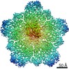
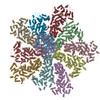
- PDB-6ogy: In situ structure of Rotavirus RNA-dependent RNA polymerase at du... -
+ Open data

- Basic information
| Entry | Database: PDB / ID: 6ogy | |||||||||||||||||||||||||||||||||||||||||||||||||||
|---|---|---|---|---|---|---|---|---|---|---|---|---|---|---|---|---|---|---|---|---|---|---|---|---|---|---|---|---|---|---|---|---|---|---|---|---|---|---|---|---|---|---|---|---|---|---|---|---|---|---|---|---|
| Title | In situ structure of Rotavirus RNA-dependent RNA polymerase at duplex-open state | |||||||||||||||||||||||||||||||||||||||||||||||||||
 Components Components |
| |||||||||||||||||||||||||||||||||||||||||||||||||||
 Keywords Keywords | viral protein/transferase/rna / RNA-dependent RNA polymerase / capsid shell protein / transcription / in situ structure / rotavirus / transcriptional factors / reovirus / VIRUS / viral protein-transferase-rna complex | |||||||||||||||||||||||||||||||||||||||||||||||||||
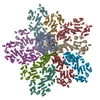
| Function / homology |  Function and homology information Function and homology informationT=2 icosahedral viral capsid / viral inner capsid / viral genome replication / virion component / viral nucleocapsid / RNA-directed RNA polymerase / nucleotide binding / RNA-directed RNA polymerase activity / DNA-templated transcription / RNA binding Similarity search - Function | |||||||||||||||||||||||||||||||||||||||||||||||||||
| Biological species |  Rotavirus A Rotavirus A | |||||||||||||||||||||||||||||||||||||||||||||||||||
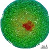
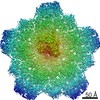
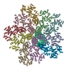
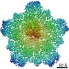
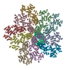
| Method | ELECTRON MICROSCOPY / single particle reconstruction / cryo EM / Resolution: 3.4 Å | |||||||||||||||||||||||||||||||||||||||||||||||||||
 Authors Authors | Ding, K. / Chang, T. / Shen, W. / Roy, P. / Zhou, Z.H. | |||||||||||||||||||||||||||||||||||||||||||||||||||
| Funding support |  United States, 8items United States, 8items
| |||||||||||||||||||||||||||||||||||||||||||||||||||
 Citation Citation | Journal: Nat Commun / Year: 2019 Title: In situ structures of rotavirus polymerase in action and mechanism of mRNA transcription and release. Authors: Ke Ding / Cristina C Celma / Xing Zhang / Thomas Chang / Wesley Shen / Ivo Atanasov / Polly Roy / Z Hong Zhou /  Abstract: Transcribing and replicating a double-stranded genome require protein modules to unwind, transcribe/replicate nucleic acid substrates, and release products. Here we present in situ cryo-electron ...Transcribing and replicating a double-stranded genome require protein modules to unwind, transcribe/replicate nucleic acid substrates, and release products. Here we present in situ cryo-electron microscopy structures of rotavirus dsRNA-dependent RNA polymerase (RdRp) in two states pertaining to transcription. In addition to the previously discovered universal "hand-shaped" polymerase core domain shared by DNA polymerases and telomerases, our results show the function of N- and C-terminal domains of RdRp: the former opens the genome duplex to isolate the template strand; the latter splits the emerging template-transcript hybrid, guides genome reannealing to form a transcription bubble, and opens a capsid shell protein (CSP) to release the transcript. These two "helicase" domains also extensively interact with CSP, which has a switchable N-terminal helix that, like cellular transcriptional factors, either inhibits or promotes RdRp activity. The in situ structures of RdRp, CSP, and RNA in action inform mechanisms of not only transcription, but also replication. | |||||||||||||||||||||||||||||||||||||||||||||||||||
| History |
|
- Structure visualization
| Movie |
Movie viewer |
|---|---|
| Structure viewer | Molecule: MolmilJmol/JSmol |
- Downloads & links
-Download
| PDBx/mmCIF format | 6ogy.cif.gz | 1.6 MB | Display | PDBx/mmCIF format |
|---|---|---|---|---|
| PDB format | pdb6ogy.ent.gz | 1.3 MB | Display | PDB format |
| PDBx/mmJSON format | 6ogy.json.gz | Tree view | PDBx/mmJSON format | |
| Others |  Other downloads Other downloads |
-Validation report
| Arichive directory | https://data.pdbj.org/pub/pdb/validation_reports/og/6ogyftp://data.pdbj.org/pub/pdb/validation_reports/og/6ogy | HTTPS FTP |
|---|
-Related structure data
| Related structure data | 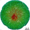 20059MC 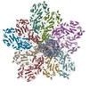 6ogzC M: map data used to model this data C: citing same article ( |
|---|---|
| Similar structure data |


-Links
 PDBj
PDBj
- Assembly
| Deposited unit | 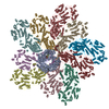
|
|---|---|
| 1 |
|
-Components
| #1: Protein | Mass: 125276.305 Da / Num. of mol.: 1 / Source method: isolated from a natural source / Source: (natural) Rotavirus A / References: UniProt: G0YZJ9, RNA-directed RNA polymerase | ||||||
|---|---|---|---|---|---|---|---|
| #2: Protein | Mass: 103425.992 Da / Num. of mol.: 10 / Source method: isolated from a natural source / Source: (natural) Rotavirus A / References: UniProt: G0YZK0#3: DNA/RNA hybrid | | Mass: 1390.855 Da / Num. of mol.: 1 / Source method: obtained synthetically / Source: (synth.) Rotavirus A#4: RNA chain | | Mass: 1239.818 Da / Num. of mol.: 1 / Source method: isolated from a natural source / Source: (natural) Rotavirus AHas protein modification | N | |
-Experimental details
-Experiment
| Experiment | Method: ELECTRON MICROSCOPY |
|---|---|
| EM experiment | Aggregation state: PARTICLE / 3D reconstruction method: single particle reconstruction |
- Sample preparation
| Component | Name: Rotavirus A / Type: VIRUS / Entity ID: all / Source: NATURAL |
|---|---|
| Molecular weight | Experimental value: NO |
| Source (natural) | Organism: Rotavirus A |
| Details of virus | Empty: NO / Enveloped: NO / Isolate: SPECIES / Type: VIRION |
| Buffer solution | pH: 7.4 |
| Specimen | Embedding applied: NO / Shadowing applied: NO / Staining applied: NO / Vitrification applied: YES |
| Vitrification | Cryogen name: ETHANE |
- Electron microscopy imaging
| Experimental equipment |  Model: Titan Krios / Image courtesy: FEI Company |
|---|---|
| Microscopy | Model: FEI TITAN KRIOS |
| Electron gun | Electron source:  FIELD EMISSION GUN / Accelerating voltage: 300 kV / Illumination mode: FLOOD BEAM FIELD EMISSION GUN / Accelerating voltage: 300 kV / Illumination mode: FLOOD BEAM |
| Electron lens | Mode: BRIGHT FIELD / Cs: 2.7 mm |
| Image recording | Electron dose: 22 e/Å2 / Detector mode: COUNTING / Film or detector model: GATAN K2 SUMMIT (4k x 4k) |
| Image scans | Width: 3838 / Height: 3710 / Movie frames/image: 25 |
- Processing
| Software | Name: PHENIX / Version: (1.14_3260: phenix.real_space_refine) / Classification: refinement | ||||||||||||||||||||||||||||||||||||
|---|---|---|---|---|---|---|---|---|---|---|---|---|---|---|---|---|---|---|---|---|---|---|---|---|---|---|---|---|---|---|---|---|---|---|---|---|---|
| EM software |
| ||||||||||||||||||||||||||||||||||||
| CTF correction | Type: PHASE FLIPPING AND AMPLITUDE CORRECTION | ||||||||||||||||||||||||||||||||||||
| Symmetry | Point symmetry: C1 (asymmetric) | ||||||||||||||||||||||||||||||||||||
| 3D reconstruction | Resolution: 3.4 Å / Resolution method: FSC 0.143 CUT-OFF / Num. of particles: 798260 / Symmetry type: POINT |
