 Movie
Movie Controller


+ Open data

- Basic information
| Entry | Database: PDB / ID: 6lvb | ||||||||||||
|---|---|---|---|---|---|---|---|---|---|---|---|---|---|
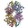
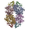
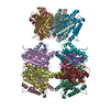
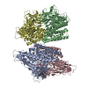
| Title | Structure of Dimethylformamidase, tetramer | ||||||||||||
 Components Components |
| ||||||||||||
 Keywords Keywords | HYDROLASE / ab polypeptide / mononuclear iron / amidohydrolase / tetramer | ||||||||||||
| Function / homology |  Function and homology information Function and homology informationN,N-dimethylformamidase / N,N-dimethylformamidase activity / metal ion binding Similarity search - Function | ||||||||||||
| Biological species |  Paracoccus sp. SSG05 (bacteria) Paracoccus sp. SSG05 (bacteria) | ||||||||||||
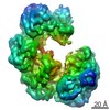
| Method | ELECTRON MICROSCOPY / single particle reconstruction / cryo EM / Resolution: 2.8 Å | ||||||||||||
 Authors Authors | Arya, C.A. / Yadav, S. / Fine, J. / Casanal, A. / Chopra, G. / Ramanathan, G. / Subramanian, R. / Vinothkumar, K.R. | ||||||||||||
| Funding support |  India, 3items India, 3items
| ||||||||||||
 Citation Citation | Journal: Angew Chem Int Ed Engl / Year: 2020 Title: A 2-Tyr-1-carboxylate Mononuclear Iron Center Forms the Active Site of a Paracoccus Dimethylformamidase. Authors: Chetan Kumar Arya / Swati Yadav / Jonathan Fine / Ana Casanal / Gaurav Chopra / Gurunath Ramanathan / Kutti R Vinothkumar / Ramaswamy Subramanian /   Abstract: N,N-dimethyl formamide (DMF) is an extensively used organic solvent but is also a potent pollutant. Certain bacterial species from genera such as Paracoccus, Pseudomonas, and Alcaligenes have evolved ...N,N-dimethyl formamide (DMF) is an extensively used organic solvent but is also a potent pollutant. Certain bacterial species from genera such as Paracoccus, Pseudomonas, and Alcaligenes have evolved to use DMF as a sole carbon and nitrogen source for growth via degradation by a dimethylformamidase (DMFase). We show that DMFase from Paracoccus sp. strain DMF is a halophilic and thermostable enzyme comprising a multimeric complex of the α β or (α β ) type. One of the three domains of the large subunit and the small subunit are hitherto undescribed protein folds of unknown evolutionary origin. The active site consists of a mononuclear iron coordinated by two Tyr side-chain phenolates and one carboxylate from Glu. The Fe ion in the active site catalyzes the hydrolytic cleavage of the amide bond in DMF. Kinetic characterization reveals that the enzyme shows cooperativity between subunits, and mutagenesis and structural data provide clues to the catalytic mechanism. #1: Journal: Prog Biophys Mol Biol / Year: 2021 Title: Comparison of CryoEM and X-ray structures of dimethylformamidase. Authors: Kutti R Vinothkumar / Chetan Kumar Arya / Gurunath Ramanathan / Ramaswamy Subramanian / Abstract: Dimethylformamidase (DMFase) catalyzes the hydrolysis of dimethylformamide, an industrial solvent, introduced into the environment by humans. Recently, we determined the structures of ...Dimethylformamidase (DMFase) catalyzes the hydrolysis of dimethylformamide, an industrial solvent, introduced into the environment by humans. Recently, we determined the structures of dimethylformamidase by electron cryo microscopy and X-ray crystallography revealing a tetrameric enzyme with a mononuclear iron at the active site. DMFase from Paracoccus sp. isolated from a waste water treatment plant around the city of Kanpur in India shows maximal activity at 54 °C and is halotolerant. The structures determined by both techniques are mostly identical and the largest difference is in a loop near the active site. This loop could play a role in co-operativity between the monomers. A number of non-protein densities are observed in the EM map, which are modelled as water molecules. Comparison of the structures determined by the two methods reveals conserved water molecules that could play a structural role. The higher stability, unusual active site and negligible activity at low temperature makes this a very good model to study enzyme mechanism by cryoEM. | ||||||||||||
| History |
|
- Structure visualization
| Movie |
Movie viewer |
|---|---|
| Structure viewer | Molecule: MolmilJmol/JSmol |
- Downloads & links
-Download
| PDBx/mmCIF format | 6lvb.cif.gz | 610.6 KB | Display | PDBx/mmCIF format |
|---|---|---|---|---|
| PDB format | pdb6lvb.ent.gz | 503.7 KB | Display | PDB format |
| PDBx/mmJSON format | 6lvb.json.gz | Tree view | PDBx/mmJSON format | |
| Others |  Other downloads Other downloads |
-Validation report
| Arichive directory | https://data.pdbj.org/pub/pdb/validation_reports/lv/6lvbftp://data.pdbj.org/pub/pdb/validation_reports/lv/6lvb | HTTPS FTP |
|---|
-Related structure data
| Related structure data | 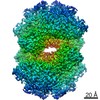 0988MC  0989C  0990C  0991C  6lvcC 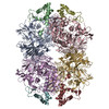 6lvdC 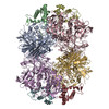 6lveC  6lvvC M: map data used to model this data C: citing same article ( |
|---|---|
| Similar structure data |


-Links
 PDBj
PDBj- Assembly
| Deposited unit | 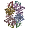
|
|---|---|
| 1 |
|
-Components
| #1: Protein | Mass: 86341.758 Da / Num. of mol.: 4 Source method: isolated from a genetically manipulated source Source: (gene. exp.) Paracoccus sp. SSG05 (bacteria) / Gene: dmfase2 / Production host: #2: Protein | Mass: 16083.823 Da / Num. of mol.: 4 Source method: isolated from a genetically manipulated source Source: (gene. exp.) Paracoccus sp. SSG05 (bacteria) / Gene: dmfase1 / Production host: #3: Chemical | ChemComp-FE /   Mass: 55.845 Da / Num. of mol.: 4 / Source method: obtained synthetically / Formula: Fe / Feature type: SUBJECT OF INVESTIGATION Mass: 55.845 Da / Num. of mol.: 4 / Source method: obtained synthetically / Formula: Fe / Feature type: SUBJECT OF INVESTIGATION#4: Water | ChemComp-HOH / |  Mass: 18.015 Da / Num. of mol.: 4 / Source method: isolated from a natural source / Formula: H2O Mass: 18.015 Da / Num. of mol.: 4 / Source method: isolated from a natural source / Formula: H2OHas ligand of interest | Y | |
|---|
-Experimental details
-Experiment
| Experiment | Method: ELECTRON MICROSCOPY |
|---|---|
| EM experiment | Aggregation state: PARTICLE / 3D reconstruction method: single particle reconstruction |
- Sample preparation
| Component | Name: Dimethylformamidase / Type: COMPLEX / Details: Tetramer, 2x(a2b2) / Entity ID: #1-#2 / Source: RECOMBINANT | ||||||||||||
|---|---|---|---|---|---|---|---|---|---|---|---|---|---|
| Molecular weight | Value: 0.4 MDa / Experimental value: NO | ||||||||||||
| Source (natural) | Organism: Paracoccus sp. SSG05 (bacteria) | ||||||||||||
| Source (recombinant) | Organism: | ||||||||||||
| Buffer solution | pH: 7.2 | ||||||||||||
| Buffer component |
| ||||||||||||
| Specimen | Conc.: 3 mg/ml / Embedding applied: NO / Shadowing applied: NO / Staining applied: NO / Vitrification applied: YES / Details: Sample at higher salt exists mostly as tetramer | ||||||||||||
| Specimen support | Details: Glow discharge was carried out in Quorum Glocube instrument at 25mA for 90 seconds. Grid material: GOLD / Grid mesh size: 300 divisions/in. / Grid type: Quantifoil R0.6/1 | ||||||||||||
| Vitrification | Instrument: FEI VITROBOT MARK IV / Cryogen name: ETHANE / Humidity: 100 % / Chamber temperature: 291 K Details: Blot force was 0 and blotting was done for 3.5 seconds |
- Electron microscopy imaging
| Experimental equipment |  Model: Titan Krios / Image courtesy: FEI Company |
|---|---|
| Microscopy | Model: FEI TITAN KRIOS |
| Electron gun | Electron source:  FIELD EMISSION GUN / Accelerating voltage: 300 kV / Illumination mode: FLOOD BEAM FIELD EMISSION GUN / Accelerating voltage: 300 kV / Illumination mode: FLOOD BEAM |
| Electron lens | Mode: BRIGHT FIELD / Nominal magnification: 75000 X / Calibrated magnification: 130841 X / Nominal defocus max: 4145.7 nm / Nominal defocus min: 931.5 nm / Calibrated defocus min: 931.5 nm / Calibrated defocus max: 4145.7 nm / Cs: 2.7 mm / C2 aperture diameter: 50 µm / Alignment procedure: COMA FREE |
| Specimen holder | Cryogen: NITROGEN / Specimen holder model: FEI TITAN KRIOS AUTOGRID HOLDER / Temperature (max): 77.7 K / Temperature (min): 77.7 K |
| Image recording | Average exposure time: 60 sec. / Electron dose: 27 e/Å2 / Detector mode: COUNTING / Film or detector model: FEI FALCON III (4k x 4k) / Num. of grids imaged: 1 / Num. of real images: 793 Details: A total of 25 frames were saved from the 60 second exposure, resulting in ~1.08 electron/frame |
| Image scans | Sampling size: 14 µm / Width: 4096 / Height: 4096 |
- Processing
| EM software |
| |||||||||||||||||||||||||||||||||||||||||||||
|---|---|---|---|---|---|---|---|---|---|---|---|---|---|---|---|---|---|---|---|---|---|---|---|---|---|---|---|---|---|---|---|---|---|---|---|---|---|---|---|---|---|---|---|---|---|---|
| CTF correction | Details: CTF correction was done with Relion, during refinement and reconstruction Type: PHASE FLIPPING AND AMPLITUDE CORRECTION | |||||||||||||||||||||||||||||||||||||||||||||
| Particle selection | Num. of particles selected: 89183 Details: Two rounds of 2D classification was performed prior to angular assignment and reconstruction | |||||||||||||||||||||||||||||||||||||||||||||
| Symmetry | Point symmetry: C2 (2 fold cyclic) | |||||||||||||||||||||||||||||||||||||||||||||
| 3D reconstruction | Resolution: 2.8 Å / Resolution method: FSC 0.143 CUT-OFF / Num. of particles: 59191 / Algorithm: BACK PROJECTION / Symmetry type: POINT | |||||||||||||||||||||||||||||||||||||||||||||
| Atomic model building | B value: 38.8 / Protocol: OTHER / Space: REAL |
