 Movie
Movie Controller


+ Open data

- Basic information
| Entry | Database: PDB / ID: 6a69 | ||||||
|---|---|---|---|---|---|---|---|
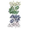
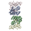
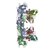
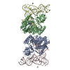
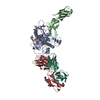
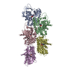
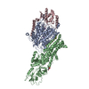
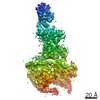
| Title | Cryo-EM structure of a P-type ATPase | ||||||
 Components Components |
| ||||||
 Keywords Keywords | STRUCTURAL PROTEIN / Membrane protein | ||||||
| Function / homology |  Function and homology information Function and homology informationcalcium ion export across plasma membrane / calcium ion transmembrane transporter activity / type 1 fibroblast growth factor receptor binding / dendrite self-avoidance / positive regulation of fibroblast growth factor receptor signaling pathway / photoreceptor ribbon synapse / regulation of vascular associated smooth muscle contraction / cell-cell adhesion mediator activity / positive regulation of long-term neuronal synaptic plasticity / P-type Ca2+ transporter ...calcium ion export across plasma membrane / calcium ion transmembrane transporter activity / type 1 fibroblast growth factor receptor binding / dendrite self-avoidance / positive regulation of fibroblast growth factor receptor signaling pathway / photoreceptor ribbon synapse / regulation of vascular associated smooth muscle contraction / cell-cell adhesion mediator activity / positive regulation of long-term neuronal synaptic plasticity / P-type Ca2+ transporter / P-type calcium transporter activity / positive regulation of calcium ion transport / GABA receptor activation / molecular function inhibitor activity / Sensory processing of sound by inner hair cells of the cochlea / negative regulation of cytokine production / Reduction of cytosolic Ca++ levels / negative regulation of cytosolic calcium ion concentration / homophilic cell-cell adhesion / Ion transport by P-type ATPases / immunological synapse / regulation of cardiac conduction / positive regulation of bone mineralization / regulation of cytosolic calcium ion concentration / Ion homeostasis / regulation of cellular response to insulin stimulus / cell adhesion molecule binding / axon guidance / positive regulation of neuron projection development / positive regulation of protein phosphorylation / regulation of blood pressure / intracellular calcium ion homeostasis / long-term synaptic potentiation / synaptic vesicle membrane / positive regulation of cytosolic calcium ion concentration / presynaptic membrane / monoatomic ion transmembrane transport / basolateral plasma membrane / calmodulin binding / postsynaptic density / axon / dendrite / glutamatergic synapse / cell surface / ATP hydrolysis activity / extracellular exosome / ATP binding / membrane / metal ion binding / plasma membrane Similarity search - Function | ||||||
| Biological species |  Homo sapiens (human) Homo sapiens (human) | ||||||
| Method | ELECTRON MICROSCOPY / single particle reconstruction / cryo EM / Resolution: 4.11 Å | ||||||
 Authors Authors | Gong, D.S. / Chi, X.M. / Ren, K. / Huang, G.X.Y. / Zhou, G.W. / Yan, N. / Lei, J.L. / Zhou, Q. | ||||||
 Citation Citation | Journal: Nat Commun / Year: 2018 Title: Structure of the human plasma membrane Ca-ATPase 1 in complex with its obligatory subunit neuroplastin. Authors: Deshun Gong / Ximin Chi / Kang Ren / Gaoxingyu Huang / Gewei Zhou / Nieng Yan / Jianlin Lei / Qiang Zhou /   Abstract: Plasma membrane Ca-ATPases (PMCAs) are key regulators of global Ca homeostasis and local intracellular Ca dynamics. Recently, Neuroplastin (NPTN) and basigin were identified as previously ...Plasma membrane Ca-ATPases (PMCAs) are key regulators of global Ca homeostasis and local intracellular Ca dynamics. Recently, Neuroplastin (NPTN) and basigin were identified as previously unrecognized obligatory subunits of PMCAs that dramatically increase the efficiency of PMCA-mediated Ca clearance. Here, we report the cryo-EM structure of human PMCA1 (hPMCA1) in complex with NPTN at a resolution of 4.1 Å for the overall structure and 3.9 Å for the transmembrane domain. The single transmembrane helix of NPTN interacts with the TM-linker and TM10 of hPMCA1. The subunits are required for the hPMCA1 functional activity. The NPTN-bound hPMCA1 closely resembles the E1-Mg structure of endo(sarco)plasmic reticulum Ca ATPase and the Ca site is exposed through a large open cytoplasmic pathway. This structure provides insight into how the subunits bind to the PMCAs and serves as an important basis for understanding the functional mechanisms of this essential calcium pump family. | ||||||
| History |
|
- Structure visualization
| Movie |
Movie viewer |
|---|---|
| Structure viewer | Molecule: MolmilJmol/JSmol |
- Downloads & links
-Download
| PDBx/mmCIF format | 6a69.cif.gz | 200 KB | Display | PDBx/mmCIF format |
|---|---|---|---|---|
| PDB format | pdb6a69.ent.gz | 149.3 KB | Display | PDB format |
| PDBx/mmJSON format | 6a69.json.gz | Tree view | PDBx/mmJSON format | |
| Others |  Other downloads Other downloads |
-Validation report
| Arichive directory | https://data.pdbj.org/pub/pdb/validation_reports/a6/6a69ftp://data.pdbj.org/pub/pdb/validation_reports/a6/6a69 | HTTPS FTP |
|---|
-Related structure data
| Related structure data |  6987MC M: map data used to model this data C: citing same article ( |
|---|---|
| Similar structure data |
-Links
 PDBj
PDBj


- Assembly
| Deposited unit | 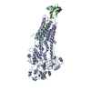
|
|---|---|
| 1 |
|
-Components
| #1: Protein | Mass: 140987.844 Da / Num. of mol.: 1 Source method: isolated from a genetically manipulated source Source: (gene. exp.) Homo sapiens (human) / Gene: ATP2B1, PMCA1 / Cell line (production host): HEK293F / Production host: Homo sapiens (human) / References: UniProt: P20020, EC: 3.6.3.8 |
|---|---|
| #2: Protein | Mass: 31328.400 Da / Num. of mol.: 1 / Source method: isolated from a natural source / Source: (natural) Homo sapiens (human) / References: UniProt: Q9Y639 |
| #3: Sugar | ChemComp-NAG /   Type: D-saccharide, beta linking / Mass: 221.208 Da / Num. of mol.: 1 Type: D-saccharide, beta linking / Mass: 221.208 Da / Num. of mol.: 1Source method: isolated from a genetically manipulated source Formula: C8H15NO6 |
| Has protein modification | Y |
| Sequence details | THIS SEQUENCE OF NPTN CORRESPOND |
-Experimental details
-Experiment
| Experiment | Method: ELECTRON MICROSCOPY |
|---|---|
| EM experiment | Aggregation state: PARTICLE / 3D reconstruction method: single particle reconstruction |
- Sample preparation
| Component | Name: complex of one PMCA1 molecular with one NPTN molecular Type: COMPLEX / Entity ID: #1-#2 / Source: RECOMBINANT |
|---|---|
| Source (natural) | Organism: Homo sapiens (human) |
| Source (recombinant) | Organism: Homo sapiens (human) |
| Buffer solution | pH: 8 |
| Specimen | Embedding applied: NO / Shadowing applied: NO / Staining applied: NO / Vitrification applied: YES |
| Vitrification | Cryogen name: ETHANE |
- Electron microscopy imaging
| Experimental equipment |  Model: Titan Krios / Image courtesy: FEI Company |
|---|---|
| Microscopy | Model: FEI TITAN KRIOS |
| Electron gun | Electron source:  FIELD EMISSION GUN / Accelerating voltage: 300 kV / Illumination mode: FLOOD BEAM FIELD EMISSION GUN / Accelerating voltage: 300 kV / Illumination mode: FLOOD BEAM |
| Electron lens | Mode: BRIGHT FIELD |
| Image recording | Electron dose: 48 e/Å2 / Film or detector model: GATAN K2 SUMMIT (4k x 4k) |
- Processing
| Software | Name: PHENIX / Version: 1.12_2829: / Classification: refinement | ||||||||||||||||||||||||||||||||||||||||
|---|---|---|---|---|---|---|---|---|---|---|---|---|---|---|---|---|---|---|---|---|---|---|---|---|---|---|---|---|---|---|---|---|---|---|---|---|---|---|---|---|---|
| EM software |
| ||||||||||||||||||||||||||||||||||||||||
| CTF correction | Type: PHASE FLIPPING AND AMPLITUDE CORRECTION | ||||||||||||||||||||||||||||||||||||||||
| Symmetry | Point symmetry: C1 (asymmetric) | ||||||||||||||||||||||||||||||||||||||||
| 3D reconstruction | Resolution: 4.11 Å / Resolution method: FSC 0.143 CUT-OFF / Num. of particles: 105188 / Symmetry type: POINT | ||||||||||||||||||||||||||||||||||||||||
| Refinement | Highest resolution: 4.11 Å | ||||||||||||||||||||||||||||||||||||||||
| Refine LS restraints |
|
