 Movie
Movie Controller

[English] 日本語
 Yorodumi
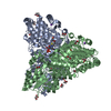
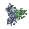
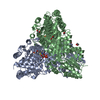
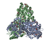
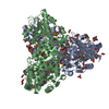
- PDB-6tj8: Escherichia coli transketolase in complex with cofactor analog 2'... -
+ Open data

- Basic information
| Entry | Database: PDB / ID: 6tj8 | ||||||
|---|---|---|---|---|---|---|---|
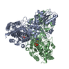
| Title | Escherichia coli transketolase in complex with cofactor analog 2'-methoxythiamine diphosphate | ||||||
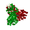
 Components Components | Transketolase 1 | ||||||
 Keywords Keywords | TRANSFERASE / THIAMIN DIPHOSPHATE / ENZYME CATALYSIS / PENTOSE PHOSPHATE PATHWAY | ||||||
| Function / homology |  Function and homology information Function and homology informationtransketolase / transketolase activity / pentose-phosphate shunt, non-oxidative branch / pentose-phosphate shunt / thiamine pyrophosphate binding / manganese ion binding / magnesium ion binding / protein homodimerization activity / cytosol Similarity search - Function | ||||||
| Biological species |  | ||||||
| Method |  X-RAY DIFFRACTION / SYNCHROTRON / MOLECULAR REPLACEMENT / Resolution: 0.921 Å X-RAY DIFFRACTION / SYNCHROTRON / MOLECULAR REPLACEMENT / Resolution: 0.921 Å | ||||||
 Authors Authors | Rabe von Pappenheim, F. / Tittmann, K. | ||||||
| Funding support |  Germany, 1items Germany, 1items
| ||||||
 Citation Citation | Journal: Nat.Chem.Biol. / Year: 2020 Title: Structural basis for antibiotic action of the B 1 antivitamin 2'-methoxy-thiamine. Authors: Rabe von Pappenheim, F. / Aldeghi, M. / Shome, B. / Begley, T. / de Groot, B.L. / Tittmann, K. | ||||||
| History |
|
- Structure visualization
| Structure viewer | Molecule: MolmilJmol/JSmol |
|---|
- Downloads & links
-Download
| PDBx/mmCIF format | 6tj8.cif.gz | 879.4 KB | Display | PDBx/mmCIF format |
|---|---|---|---|---|
| PDB format | pdb6tj8.ent.gz | 739.9 KB | Display | PDB format |
| PDBx/mmJSON format | 6tj8.json.gz | Tree view | PDBx/mmJSON format | |
| Others |  Other downloads Other downloads |
-Validation report
| Summary document | 6tj8_validation.pdf.gz | 1.4 MB | Display | wwPDB validaton report |
|---|---|---|---|---|
| Full document | 6tj8_full_validation.pdf.gz | 1.4 MB | Display | |
| Data in XML | 6tj8_validation.xml.gz | 63.2 KB | Display | |
| Data in CIF | 6tj8_validation.cif.gz | 101.8 KB | Display | |
| Arichive directory | https://data.pdbj.org/pub/pdb/validation_reports/tj/6tj8ftp://data.pdbj.org/pub/pdb/validation_reports/tj/6tj8 | HTTPS FTP |
-Related structure data
| Related structure data |  6tj9C  1qgdS S: Starting model for refinement C: citing same article ( |
|---|---|
| Similar structure data |

-Links
 PDBj
PDBj


- Assembly
| Deposited unit | 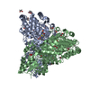
| ||||||||
|---|---|---|---|---|---|---|---|---|---|
| 1 |
| ||||||||
| Unit cell |
|
-Components
| #1: Protein | Mass: 73121.508 Da / Num. of mol.: 2 Source method: isolated from a genetically manipulated source Source: (gene. exp.) Strain: K12 / Gene: tktA, tkt, b2935, JW5478 / Production host: #2: Chemical | 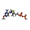  Mass: 441.314 Da / Num. of mol.: 2 / Source method: obtained synthetically / Formula: C12H19N4O8P2S / Feature type: SUBJECT OF INVESTIGATION Mass: 441.314 Da / Num. of mol.: 2 / Source method: obtained synthetically / Formula: C12H19N4O8P2S / Feature type: SUBJECT OF INVESTIGATION#3: Chemical |   Mass: 40.078 Da / Num. of mol.: 2 / Source method: obtained synthetically / Formula: Ca Mass: 40.078 Da / Num. of mol.: 2 / Source method: obtained synthetically / Formula: Ca#4: Chemical | ChemComp-EDO /   Mass: 62.068 Da / Num. of mol.: 33 / Source method: obtained synthetically / Formula: C2H6O2 Mass: 62.068 Da / Num. of mol.: 33 / Source method: obtained synthetically / Formula: C2H6O2#5: Water | ChemComp-HOH / |  Mass: 18.015 Da / Num. of mol.: 1589 / Source method: isolated from a natural source / Formula: H2O Mass: 18.015 Da / Num. of mol.: 1589 / Source method: isolated from a natural source / Formula: H2OHas ligand of interest | Y | |
|---|
-Experimental details
-Experiment
| Experiment | Method: X-RAY DIFFRACTION / Number of used crystals: 1 |
|---|
- Sample preparation
| Crystal | Density Matthews: 2.08 Å3/Da / Density % sol: 40.81 % |
|---|---|
| Crystal grow | Temperature: 279.15 K / Method: vapor diffusion, hanging drop / pH: 7.9 / Details: PEG6000, Glycerol, Glycyl-Glycine |
-Data collection
| Diffraction | Mean temperature: 100 K / Serial crystal experiment: N |
|---|---|
| Diffraction source | Source: SYNCHROTRON / Site: PETRA III, EMBL c/o DESY / Beamline: P14 (MX2) / Wavelength: 1 Å |
| Detector | Type: DECTRIS PILATUS 6M-F / Detector: PIXEL / Date: Jun 2, 2016 |
| Radiation | Protocol: SINGLE WAVELENGTH / Monochromatic (M) / Laue (L): M / Scattering type: x-ray |
| Radiation wavelength | Wavelength: 1 Å / Relative weight: 1 |
| Reflection | Resolution: 0.92→47.3 Å / Num. obs: 780617 / % possible obs: 93.8 % / Redundancy: 4.2 % / Biso Wilson estimate: 7.62 Å2 / CC1/2: 0.994 / Rrim(I) all: 0.095 / Net I/σ(I): 9.73 |
| Reflection shell | Resolution: 0.92→0.95 Å / Redundancy: 2.9 % / Num. unique obs: 46733 / CC1/2: 0.67 / Rrim(I) all: 0.061 / % possible all: 61.6 |
- Processing
| Software |
| ||||||||||||||||||||||||||||||||||||||||||||||||||||||||||||||||||||||||||||||||||||||||||||||||||||||||||||||||||||||||||||||||||||||||||||||||||||||||||||||||||||||||||||||||||||||||||
|---|---|---|---|---|---|---|---|---|---|---|---|---|---|---|---|---|---|---|---|---|---|---|---|---|---|---|---|---|---|---|---|---|---|---|---|---|---|---|---|---|---|---|---|---|---|---|---|---|---|---|---|---|---|---|---|---|---|---|---|---|---|---|---|---|---|---|---|---|---|---|---|---|---|---|---|---|---|---|---|---|---|---|---|---|---|---|---|---|---|---|---|---|---|---|---|---|---|---|---|---|---|---|---|---|---|---|---|---|---|---|---|---|---|---|---|---|---|---|---|---|---|---|---|---|---|---|---|---|---|---|---|---|---|---|---|---|---|---|---|---|---|---|---|---|---|---|---|---|---|---|---|---|---|---|---|---|---|---|---|---|---|---|---|---|---|---|---|---|---|---|---|---|---|---|---|---|---|---|---|---|---|---|---|---|---|---|---|
| Refinement | Method to determine structure: MOLECULAR REPLACEMENT Starting model: 1QGD Resolution: 0.921→47.285 Å / SU ML: 0.05 / Cross valid method: THROUGHOUT / σ(F): 1.34 / Phase error: 8.39 / Stereochemistry target values: ML
| ||||||||||||||||||||||||||||||||||||||||||||||||||||||||||||||||||||||||||||||||||||||||||||||||||||||||||||||||||||||||||||||||||||||||||||||||||||||||||||||||||||||||||||||||||||||||||
| Solvent computation | Shrinkage radii: 1.3 Å / VDW probe radii: 1.4 Å / Solvent model: FLAT BULK SOLVENT MODEL | ||||||||||||||||||||||||||||||||||||||||||||||||||||||||||||||||||||||||||||||||||||||||||||||||||||||||||||||||||||||||||||||||||||||||||||||||||||||||||||||||||||||||||||||||||||||||||
| Displacement parameters | Biso max: 48.08 Å2 / Biso mean: 10.5689 Å2 / Biso min: 2.75 Å2 | ||||||||||||||||||||||||||||||||||||||||||||||||||||||||||||||||||||||||||||||||||||||||||||||||||||||||||||||||||||||||||||||||||||||||||||||||||||||||||||||||||||||||||||||||||||||||||
| Refinement step | Cycle: final / Resolution: 0.921→47.285 Å
| ||||||||||||||||||||||||||||||||||||||||||||||||||||||||||||||||||||||||||||||||||||||||||||||||||||||||||||||||||||||||||||||||||||||||||||||||||||||||||||||||||||||||||||||||||||||||||
| LS refinement shell | Refine-ID: X-RAY DIFFRACTION / Rfactor Rfree error: 0
|
