[English] 日本語
 Yorodumi
- PDB-6s7u: Fumarate hydratase of Mycobacterium tuberculosis in complex with ... -
+ Open data
- Basic information
| Entry | Database: PDB / ID: 6s7u | ||||||
|---|---|---|---|---|---|---|---|
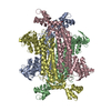
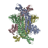
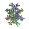
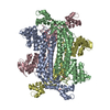
| Title | Fumarate hydratase of Mycobacterium tuberculosis in complex with formate and allosteric modulator N-(5-(Azepan-1-ylsulfonyl)-2-methoxyphenyl)-2-(1H-indol-3-yl)acetamide | ||||||
 Components Components | Fumarate hydratase class II | ||||||
 Keywords Keywords | LYASE / Fumarate hydratase / Fumarase | ||||||
| Function / homology |  Function and homology information Function and homology informationfumarate hydratase activity / fumarate hydratase / fumarate metabolic process / tricarboxylic acid cycle / peptidoglycan-based cell wall / extracellular region / plasma membrane / cytosol / cytoplasm Similarity search - Function | ||||||
| Biological species |  | ||||||
| Method |  X-RAY DIFFRACTION / X-RAY DIFFRACTION /  SYNCHROTRON / SYNCHROTRON /  MOLECULAR REPLACEMENT / MOLECULAR REPLACEMENT /  molecular replacement / Resolution: 1.48 Å molecular replacement / Resolution: 1.48 Å | ||||||
 Authors Authors | Whitehouse, A.J. / Libardo, M.D. / Kasbekar, M. / Brear, P. / Fischer, G. / Thomas, C.J. / Barry, C.E. / Boshoff, H.I. / Coyne, A.G. / Abell, C. | ||||||
| Funding support |  United Kingdom, 1items United Kingdom, 1items
| ||||||
 Citation Citation |  Journal: J.Med.Chem. / Year: 2019 Journal: J.Med.Chem. / Year: 2019Title: Targeting of Fumarate Hydratase fromMycobacterium tuberculosisUsing Allosteric Inhibitors with a Dimeric-Binding Mode. Authors: Whitehouse, A.J. / Libardo, M.D.J. / Kasbekar, M. / Brear, P.D. / Fischer, G. / Thomas, C.J. / Barry 3rd, C.E. / Boshoff, H.I.M. / Coyne, A.G. / Abell, C. | ||||||
| History |
|
- Structure visualization
| Structure viewer | Molecule:  Molmil Molmil Jmol/JSmol Jmol/JSmol |
|---|
- Downloads & links
- Download
| PDBx/mmCIF format |  6s7u.cif.gz 6s7u.cif.gz | 390.1 KB | Display |  PDBx/mmCIF format PDBx/mmCIF format |
|---|---|---|---|---|
| PDB format |  pdb6s7u.ent.gz pdb6s7u.ent.gz | 315.1 KB | Display |  PDB format PDB format |
| PDBx/mmJSON format |  6s7u.json.gz 6s7u.json.gz | Tree view |  PDBx/mmJSON format PDBx/mmJSON format | |
| Others |  Other downloads Other downloads |
-Validation report
| Arichive directory |  https://data.pdbj.org/pub/pdb/validation_reports/s7/6s7u https://data.pdbj.org/pub/pdb/validation_reports/s7/6s7u ftp://data.pdbj.org/pub/pdb/validation_reports/s7/6s7u ftp://data.pdbj.org/pub/pdb/validation_reports/s7/6s7u | HTTPS FTP |
|---|
-Related structure data
| Related structure data |  6s43SC  6s7kC  6s7sC  6s7wC  6s7zC  6s88C S: Starting model for refinement C: citing same article ( |
|---|---|
| Similar structure data |
- Links
Links
- Assembly
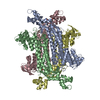
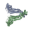
| Deposited unit | 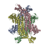
| ||||||||
|---|---|---|---|---|---|---|---|---|---|
| 1 |
| ||||||||
| Unit cell |
| ||||||||
| Components on special symmetry positions |
|
- Components
| #1: Protein | Mass: 50191.805 Da / Num. of mol.: 4 Source method: isolated from a genetically manipulated source Source: (gene. exp.)  Gene: fum, fumC, DSI35_09545, ERS007663_02818, ERS007679_02635, ERS007688_02307, ERS007722_03256, ERS023446_00966, ERS024276_01230, ERS027646_00759, ERS027653_00188, ERS027654_00515, ERS027659_00384, ...Gene: fum, fumC, DSI35_09545, ERS007663_02818, ERS007679_02635, ERS007688_02307, ERS007722_03256, ERS023446_00966, ERS024276_01230, ERS027646_00759, ERS027653_00188, ERS027654_00515, ERS027659_00384, ERS027661_00671, ERS027666_02275, ERS124361_03161, SAMEA2682864_03539, SAMEA2683035_02636 Plasmid: pNAN / Production host:  References: UniProt: A0A045IXZ8, UniProt: P9WN93*PLUS, fumarate hydratase #2: Chemical | #3: Chemical | ChemComp-FMT / | #4: Chemical | #5: Water | ChemComp-HOH / | Has ligand of interest | Y | |
|---|
-Experimental details
-Experiment
| Experiment | Method:  X-RAY DIFFRACTION / Number of used crystals: 1 X-RAY DIFFRACTION / Number of used crystals: 1 |
|---|
- Sample preparation
| Crystal | Density Matthews: 2.68 Å3/Da / Density % sol: 54.14 % / Mosaicity: 0.07 ° |
|---|---|
| Crystal grow | Temperature: 292 K / Method: vapor diffusion, sitting drop / pH: 8 / Details: NaCl, Tris, TCEP, PEG3350, DMSO, magnesium formate |
-Data collection
| Diffraction | Mean temperature: 100 K / Serial crystal experiment: N | |||||||||||||||||||||||||||
|---|---|---|---|---|---|---|---|---|---|---|---|---|---|---|---|---|---|---|---|---|---|---|---|---|---|---|---|---|
| Diffraction source | Source:  SYNCHROTRON / Site: SYNCHROTRON / Site:  Diamond Diamond  / Beamline: I03 / Wavelength: 0.9763 Å / Beamline: I03 / Wavelength: 0.9763 Å | |||||||||||||||||||||||||||
| Detector | Type: DECTRIS PILATUS3 6M / Detector: PIXEL / Date: Sep 24, 2018 | |||||||||||||||||||||||||||
| Radiation | Protocol: SINGLE WAVELENGTH / Monochromatic (M) / Laue (L): M / Scattering type: x-ray | |||||||||||||||||||||||||||
| Radiation wavelength | Wavelength: 0.9763 Å / Relative weight: 1 | |||||||||||||||||||||||||||
| Reflection | Resolution: 1.48→121.2 Å / Num. obs: 324139 / % possible obs: 96.2 % / Redundancy: 7.4 % / CC1/2: 0.998 / Rmerge(I) obs: 0.141 / Rpim(I) all: 0.055 / Rrim(I) all: 0.151 / Net I/σ(I): 8.4 / Num. measured all: 2395184 / Scaling rejects: 77 | |||||||||||||||||||||||||||
| Reflection shell | Diffraction-ID: 1 / % possible all: 99.8
|
-Phasing
| Phasing | Method:  molecular replacement molecular replacement |
|---|
- Processing
| Software |
| ||||||||||||||||||||||||
|---|---|---|---|---|---|---|---|---|---|---|---|---|---|---|---|---|---|---|---|---|---|---|---|---|---|
| Refinement | Method to determine structure:  MOLECULAR REPLACEMENT MOLECULAR REPLACEMENTStarting model: 6S43 Resolution: 1.48→121.2 Å / Cor.coef. Fo:Fc: 0.971 / Cor.coef. Fo:Fc free: 0.964 / SU B: 0.001 / SU ML: 0 / Cross valid method: THROUGHOUT / σ(F): 0 / ESU R: 0.06 / ESU R Free: 0.068 Details: HYDROGENS HAVE BEEN ADDED IN THE RIDING POSITIONS U VALUES : REFINED INDIVIDUALLY
| ||||||||||||||||||||||||
| Solvent computation | Ion probe radii: 0.8 Å / Shrinkage radii: 0.8 Å / VDW probe radii: 1.2 Å | ||||||||||||||||||||||||
| Displacement parameters | Biso max: 140.43 Å2 / Biso mean: 24.349 Å2 / Biso min: 5.84 Å2
| ||||||||||||||||||||||||
| Refinement step | Cycle: final / Resolution: 1.48→121.2 Å
| ||||||||||||||||||||||||
| LS refinement shell | Resolution: 1.48→1.516 Å / Rfactor Rfree error: 0
|
 Movie
Movie Controller




 PDBj
PDBj


