 Movie
Movie Controller

[English] 日本語
 Yorodumi
- PDB-6rbs: Crystal structure of NAD kinase 1 from Listeria monocytogenes in ... -
+ Open data

- Basic information
| Entry | Database: PDB / ID: 6rbs | ||||||
|---|---|---|---|---|---|---|---|
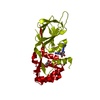
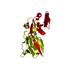
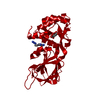
| Title | Crystal structure of NAD kinase 1 from Listeria monocytogenes in complexe with an adenine derivative | ||||||
 Components Components | NAD kinase 1 | ||||||
 Keywords Keywords | TRANSFERASE / tetrameric NAD kinase | ||||||
| Function / homology |  Function and homology information Function and homology informationNAD+ kinase / NAD+ kinase activity / NADP biosynthetic process / NAD metabolic process / NAD binding / ATP binding / metal ion binding / cytoplasm Similarity search - Function | ||||||
| Biological species |  Listeria monocytogenes serovar 1/2a (bacteria) Listeria monocytogenes serovar 1/2a (bacteria) | ||||||
| Method |  X-RAY DIFFRACTION / SYNCHROTRON / MOLECULAR REPLACEMENT / Resolution: 2.324 Å X-RAY DIFFRACTION / SYNCHROTRON / MOLECULAR REPLACEMENT / Resolution: 2.324 Å | ||||||
 Authors Authors | Gelin, M. / Labesse, G. | ||||||
 Citation Citation | Journal: Acs Infect Dis. / Year: 2020 Title: From Substrate to Fragments to Inhibitor ActiveIn VivoagainstStaphylococcus aureus. Authors: Gelin, M. / Paoletti, J. / Nahori, M.A. / Huteau, V. / Leseigneur, C. / Jouvion, G. / Dugue, L. / Clement, D. / Pons, J.L. / Assairi, L. / Pochet, S. / Labesse, G. / Dussurget, O. | ||||||
| History |
|
- Structure visualization
| Structure viewer | Molecule: MolmilJmol/JSmol |
|---|
- Downloads & links
-Download
| PDBx/mmCIF format | 6rbs.cif.gz | 119.1 KB | Display | PDBx/mmCIF format |
|---|---|---|---|---|
| PDB format | pdb6rbs.ent.gz | 91.6 KB | Display | PDB format |
| PDBx/mmJSON format | 6rbs.json.gz | Tree view | PDBx/mmJSON format | |
| Others |  Other downloads Other downloads |
-Validation report
| Summary document | 6rbs_validation.pdf.gz | 715.8 KB | Display | wwPDB validaton report |
|---|---|---|---|---|
| Full document | 6rbs_full_validation.pdf.gz | 717 KB | Display | |
| Data in XML | 6rbs_validation.xml.gz | 11.9 KB | Display | |
| Data in CIF | 6rbs_validation.cif.gz | 15.2 KB | Display | |
| Arichive directory | https://data.pdbj.org/pub/pdb/validation_reports/rb/6rbsftp://data.pdbj.org/pub/pdb/validation_reports/rb/6rbs | HTTPS FTP |
-Related structure data
| Related structure data |  6rboC  6rbpC  6rbqC  6rbrC  6rbtC  6rbuC  6rbvC  6rbwC  6rbxC  6rbyC 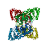 6rbzC  6rc0C  6rc1C  6rc2C  6rc3C  6rc4C 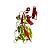 6rc5C 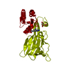 6rc6C  6rg6C  6rg7C  6rg8C  6rg9C 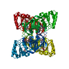 6rgaC  6rgbC  6rgcC  6rgdC  6rr2C C: citing same article ( |
|---|---|
| Similar structure data |


-Links
 PDBj
PDBj- Assembly
| Deposited unit | 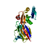
| ||||||||
|---|---|---|---|---|---|---|---|---|---|
| 1 | 
| ||||||||
| Unit cell |
|
-Components
| #1: Protein | Mass: 31045.279 Da / Num. of mol.: 1 Source method: isolated from a genetically manipulated source Source: (gene. exp.) Listeria monocytogenes serovar 1/2a (strain ATCC BAA-679 / EGD-e) (bacteria)Gene: nadK1, lmo0968 / Production host: |
|---|---|
| #2: Chemical | ChemComp-CIT /   Mass: 192.124 Da / Num. of mol.: 1 / Source method: obtained synthetically / Formula: C6H8O7 Mass: 192.124 Da / Num. of mol.: 1 / Source method: obtained synthetically / Formula: C6H8O7 |
| #3: Chemical | ChemComp-JXT /   Mass: 280.124 Da / Num. of mol.: 1 / Source method: obtained synthetically / Formula: C10H10BrN5 Mass: 280.124 Da / Num. of mol.: 1 / Source method: obtained synthetically / Formula: C10H10BrN5 |
| #4: Water | ChemComp-HOH /  Mass: 18.015 Da / Num. of mol.: 23 / Source method: isolated from a natural source / Formula: H2O Mass: 18.015 Da / Num. of mol.: 23 / Source method: isolated from a natural source / Formula: H2O |
-Experimental details
-Experiment
| Experiment | Method: X-RAY DIFFRACTION / Number of used crystals: 1 |
|---|
- Sample preparation
| Crystal | Density Matthews: 2.28 Å3/Da / Density % sol: 46.08 % |
|---|---|
| Crystal grow | Temperature: 291.15 K / Method: evaporation / pH: 5 Details: 30 mM NaBr, 220 mM Kcitrate, glycerol 6%, 15-16% w/v PEG400 |
-Data collection
| Diffraction | Mean temperature: 100 K / Serial crystal experiment: N | ||||||||||||||||||||||||||||||
|---|---|---|---|---|---|---|---|---|---|---|---|---|---|---|---|---|---|---|---|---|---|---|---|---|---|---|---|---|---|---|---|
| Diffraction source | Source: SYNCHROTRON / Site: ESRF  / Beamline: MASSIF-3 / Wavelength: 0.9677 Å / Beamline: MASSIF-3 / Wavelength: 0.9677 Å | ||||||||||||||||||||||||||||||
| Detector | Type: DECTRIS EIGER X 4M / Detector: PIXEL / Date: Jun 7, 2018 | ||||||||||||||||||||||||||||||
| Radiation | Protocol: SINGLE WAVELENGTH / Monochromatic (M) / Laue (L): M / Scattering type: x-ray | ||||||||||||||||||||||||||||||
| Radiation wavelength | Wavelength: 0.9677 Å / Relative weight: 1 | ||||||||||||||||||||||||||||||
| Reflection | Resolution: 1.822→64.277 Å / Num. obs: 13646 / % possible obs: 92.4 % / Redundancy: 5.4 % / Biso Wilson estimate: 47.41 Å2 / CC1/2: 0.999 / Rmerge(I) obs: 0.068 / Rpim(I) all: 0.032 / Rrim(I) all: 0.076 / Net I/σ(I): 14.2 / Num. measured all: 73857 | ||||||||||||||||||||||||||||||
| Reflection shell | Diffraction-ID: 1
|
- Processing
| Software |
| ||||||||||||||||||||||||||||||||||||||||||||||||||||||||||||||||||||||||||||||||||||||||||||||||
|---|---|---|---|---|---|---|---|---|---|---|---|---|---|---|---|---|---|---|---|---|---|---|---|---|---|---|---|---|---|---|---|---|---|---|---|---|---|---|---|---|---|---|---|---|---|---|---|---|---|---|---|---|---|---|---|---|---|---|---|---|---|---|---|---|---|---|---|---|---|---|---|---|---|---|---|---|---|---|---|---|---|---|---|---|---|---|---|---|---|---|---|---|---|---|---|---|---|
| Refinement | Method to determine structure: MOLECULAR REPLACEMENT / Resolution: 2.324→35.075 Å / SU ML: 0.34 / Cross valid method: THROUGHOUT / σ(F): 1.35 / Phase error: 29.92
| ||||||||||||||||||||||||||||||||||||||||||||||||||||||||||||||||||||||||||||||||||||||||||||||||
| Solvent computation | Shrinkage radii: 0.9 Å / VDW probe radii: 1.11 Å | ||||||||||||||||||||||||||||||||||||||||||||||||||||||||||||||||||||||||||||||||||||||||||||||||
| Displacement parameters | Biso max: 133.06 Å2 / Biso mean: 58.0999 Å2 / Biso min: 30.13 Å2 | ||||||||||||||||||||||||||||||||||||||||||||||||||||||||||||||||||||||||||||||||||||||||||||||||
| Refinement step | Cycle: final / Resolution: 2.324→35.075 Å
| ||||||||||||||||||||||||||||||||||||||||||||||||||||||||||||||||||||||||||||||||||||||||||||||||
| LS refinement shell | Refine-ID: X-RAY DIFFRACTION / Rfactor Rfree error: 0 / Total num. of bins used: 4
| ||||||||||||||||||||||||||||||||||||||||||||||||||||||||||||||||||||||||||||||||||||||||||||||||
| Refinement TLS params. | S33: 0 Å ° / Method: refined / Refine-ID: X-RAY DIFFRACTION
| ||||||||||||||||||||||||||||||||||||||||||||||||||||||||||||||||||||||||||||||||||||||||||||||||
| Refinement TLS group |
|
