 Movie
Movie Controller

[English] 日本語
 Yorodumi
- PDB-6o8v: The structure of lipase from Thermomyces Lanuginosa in complex wi... -
+ Open data

- Basic information
| Entry | Database: PDB / ID: 6o8v | ||||||
|---|---|---|---|---|---|---|---|
| Title | The structure of lipase from Thermomyces Lanuginosa in complex with 1,3 diacylglycerol: Rhombohedral crystal form | ||||||
 Components Components | Lipase | ||||||
 Keywords Keywords | HYDROLASE / trimer / esterase / lipids / lipase / substrate complex / catalysis / acyl intermediate / LIPID BINDING PROTEIN | ||||||
| Function / homology |  Function and homology information Function and homology informationtriacylglycerol lipase / triacylglycerol lipase activity / lipid catabolic process Similarity search - Function | ||||||
| Biological species |   Thermomyces lanuginosus (fungus) Thermomyces lanuginosus (fungus) | ||||||
| Method |  X-RAY DIFFRACTION / SYNCHROTRON / MOLECULAR REPLACEMENT / Resolution: 1.3 Å X-RAY DIFFRACTION / SYNCHROTRON / MOLECULAR REPLACEMENT / Resolution: 1.3 Å | ||||||
 Authors Authors | McPherson, A. | ||||||
| Funding support | 1items
| ||||||
 Citation Citation | Journal: Curr Enzym Inhib / Year: 2020 Title: The Crystal Structures of Thermomyces (Humicola) Lanuginosa Lipase in Complex with Enzymatic Reactants Authors: McPherson , A. / Larson , B.S. / Kalasky , A. | ||||||
| History |
|
- Structure visualization
| Structure viewer | Molecule: MolmilJmol/JSmol |
|---|
- Downloads & links
-Download
| PDBx/mmCIF format | 6o8v.cif.gz | 219.4 KB | Display | PDBx/mmCIF format |
|---|---|---|---|---|
| PDB format | pdb6o8v.ent.gz | 146.3 KB | Display | PDB format |
| PDBx/mmJSON format | 6o8v.json.gz | Tree view | PDBx/mmJSON format | |
| Others |  Other downloads Other downloads |
-Validation report
| Arichive directory | https://data.pdbj.org/pub/pdb/validation_reports/o8/6o8vftp://data.pdbj.org/pub/pdb/validation_reports/o8/6o8v | HTTPS FTP |
|---|
-Related structure data
| Related structure data |  6o9fC 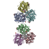 6oszC 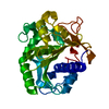 1tibS S: Starting model for refinement C: citing same article ( |
|---|---|
| Similar structure data |
-Links
 PDBj
PDBj- Assembly
| Deposited unit | 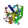
| ||||||||||||||||||
|---|---|---|---|---|---|---|---|---|---|---|---|---|---|---|---|---|---|---|---|
| 1 | 
| ||||||||||||||||||
| Unit cell |
| ||||||||||||||||||
| Components on special symmetry positions |
|
-Components
-Protein / Sugars , 2 types, 2 molecules A
| #1: Protein | Mass: 31836.459 Da / Num. of mol.: 1 Source method: isolated from a genetically manipulated source Source: (gene. exp.) Thermomyces lanuginosus (fungus) / Gene: LIP / Production host:  Thermoactinomyces vulgaris (bacteria) / References: UniProt: O59952, triacylglycerol lipase Thermoactinomyces vulgaris (bacteria) / References: UniProt: O59952, triacylglycerol lipase |
|---|---|
| #2: Sugar | ChemComp-NAG /  Type: D-saccharide, beta linking / Mass: 221.208 Da / Num. of mol.: 1 Type: D-saccharide, beta linking / Mass: 221.208 Da / Num. of mol.: 1Source method: isolated from a genetically manipulated source Formula: C8H15NO6 / Feature type: SUBJECT OF INVESTIGATION |
-Non-polymers , 4 types, 412 molecules 


| #3: Chemical | ChemComp-PG4 /  Mass: 194.226 Da / Num. of mol.: 1 / Source method: obtained synthetically / Formula: C8H18O5 / Comment: precipitant*YM Mass: 194.226 Da / Num. of mol.: 1 / Source method: obtained synthetically / Formula: C8H18O5 / Comment: precipitant*YM | ||||
|---|---|---|---|---|---|
| #4: Chemical | ChemComp-PO4 /  Mass: 94.971 Da / Num. of mol.: 5 / Source method: obtained synthetically / Formula: PO4 Mass: 94.971 Da / Num. of mol.: 5 / Source method: obtained synthetically / Formula: PO4#5: Chemical | ChemComp-LTV / |  Mass: 723.204 Da / Num. of mol.: 1 / Source method: obtained synthetically / Formula: C46H90O5 / Feature type: SUBJECT OF INVESTIGATION Mass: 723.204 Da / Num. of mol.: 1 / Source method: obtained synthetically / Formula: C46H90O5 / Feature type: SUBJECT OF INVESTIGATION#6: Water | ChemComp-HOH / | Mass: 18.015 Da / Num. of mol.: 405 / Source method: isolated from a natural source / Formula: H2O |
-Details
| Has ligand of interest | Y |
|---|---|
| Has protein modification | Y |
-Experimental details
-Experiment
| Experiment | Method: X-RAY DIFFRACTION / Number of used crystals: 1 |
|---|
- Sample preparation
| Crystal | Density Matthews: 2.13 Å3/Da / Density % sol: 42.23 % / Description: thin needles of indeterminate cross section |
|---|---|
| Crystal grow | Temperature: 298 K / Method: vapor diffusion, sitting drop / pH: 6.5 Details: From a filtered, crude nutrient broth containing 30 - 50 mg/ml lipase plus 25% PEG 3350 plus 0.10 M MES buffer at room temperature PH range: 6.0 - 7.5 |
-Data collection
| Diffraction | Mean temperature: 173 K / Ambient temp details: flash frozen in cryostream / Serial crystal experiment: N |
|---|---|
| Diffraction source | Source: SYNCHROTRON / Site: ALS  / Beamline: 8.3.1 / Wavelength: 1.03 Å / Beamline: 8.3.1 / Wavelength: 1.03 Å |
| Detector | Type: DECTRIS PILATUS 300K / Detector: PIXEL / Date: Feb 15, 2018 |
| Radiation | Protocol: SINGLE WAVELENGTH / Monochromatic (M) / Laue (L): M / Scattering type: x-ray |
| Radiation wavelength | Wavelength: 1.03 Å / Relative weight: 1 |
| Reflection | Resolution: 1.3→100 Å / Num. obs: 58358 / % possible obs: 88 % / Redundancy: 31 % / Biso Wilson estimate: 13.9 Å2 / CC1/2: 1 / Rmerge(I) obs: 0.049 / Rpim(I) all: 0.01 / Rrim(I) all: 0.05 / Rsym value: 0.046 / Net I/av σ(I): 42.7 / Net I/σ(I): 42.7 |
| Reflection shell | Resolution: 1.3→1.33 Å / Redundancy: 4.2 % / Rmerge(I) obs: 0.36 / Mean I/σ(I) obs: 2.6 / Num. unique obs: 463 / CC1/2: 0.81 / Rpim(I) all: 0.205 / Rrim(I) all: 0.461 / Rsym value: 0.34 / % possible all: 14.4 |
- Processing
| Software |
| |||||||||||||||||||||||||||||||||||||||||||||||||||||||||||||||||||||||||||||||||||||||||||||||||||||||||||||||||||||||||||||||||||||||||||||||||||
|---|---|---|---|---|---|---|---|---|---|---|---|---|---|---|---|---|---|---|---|---|---|---|---|---|---|---|---|---|---|---|---|---|---|---|---|---|---|---|---|---|---|---|---|---|---|---|---|---|---|---|---|---|---|---|---|---|---|---|---|---|---|---|---|---|---|---|---|---|---|---|---|---|---|---|---|---|---|---|---|---|---|---|---|---|---|---|---|---|---|---|---|---|---|---|---|---|---|---|---|---|---|---|---|---|---|---|---|---|---|---|---|---|---|---|---|---|---|---|---|---|---|---|---|---|---|---|---|---|---|---|---|---|---|---|---|---|---|---|---|---|---|---|---|---|---|---|---|---|
| Refinement | Method to determine structure: MOLECULAR REPLACEMENT Starting model: 1tib Resolution: 1.3→80.52 Å / SU ML: 0.0934 / Cross valid method: FREE R-VALUE / σ(F): 1.37 / Phase error: 13.2256 Stereochemistry target values: GeoStd + Monomer Library + CDL v1.2
| |||||||||||||||||||||||||||||||||||||||||||||||||||||||||||||||||||||||||||||||||||||||||||||||||||||||||||||||||||||||||||||||||||||||||||||||||||
| Solvent computation | Shrinkage radii: 0.9 Å / VDW probe radii: 1.11 Å / Solvent model: FLAT BULK SOLVENT MODEL | |||||||||||||||||||||||||||||||||||||||||||||||||||||||||||||||||||||||||||||||||||||||||||||||||||||||||||||||||||||||||||||||||||||||||||||||||||
| Displacement parameters | Biso mean: 20.61 Å2 | |||||||||||||||||||||||||||||||||||||||||||||||||||||||||||||||||||||||||||||||||||||||||||||||||||||||||||||||||||||||||||||||||||||||||||||||||||
| Refinement step | Cycle: LAST / Resolution: 1.3→80.52 Å
| |||||||||||||||||||||||||||||||||||||||||||||||||||||||||||||||||||||||||||||||||||||||||||||||||||||||||||||||||||||||||||||||||||||||||||||||||||
| Refine LS restraints |
| |||||||||||||||||||||||||||||||||||||||||||||||||||||||||||||||||||||||||||||||||||||||||||||||||||||||||||||||||||||||||||||||||||||||||||||||||||
| LS refinement shell |
|
