| 登録情報 | データベース: PDB / ID: 6ilz
|
|---|
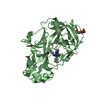
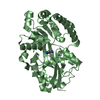
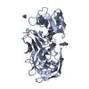
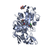
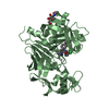
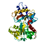
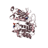
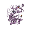
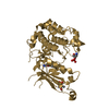
| タイトル | Crystal structure of PKCiota in complex with inhibitor |
|---|
 要素 要素 | Protein kinase C iota type |
|---|
 キーワード キーワード | TRANSFERASE / Kinase / Atypical kinase / phosphorylation / inhibitor / PKCiota / iota type / kinase domain |
|---|
| 機能・相同性 |  機能・相同性情報 機能・相同性情報
diacylglycerol-dependent, calcium-independent serine/threonine kinase activity / Golgi vesicle budding / PAR polarity complex / Tight junction interactions / protein kinase C / establishment of apical/basal cell polarity / diacylglycerol-dependent serine/threonine kinase activity / negative regulation of glial cell apoptotic process / eye photoreceptor cell development / Schmidt-Lanterman incisure ...diacylglycerol-dependent, calcium-independent serine/threonine kinase activity / Golgi vesicle budding / PAR polarity complex / Tight junction interactions / protein kinase C / establishment of apical/basal cell polarity / diacylglycerol-dependent serine/threonine kinase activity / negative regulation of glial cell apoptotic process / eye photoreceptor cell development / Schmidt-Lanterman incisure / establishment or maintenance of epithelial cell apical/basal polarity / membrane organization / cellular response to chemical stress / cell-cell junction organization / protein targeting to membrane / tight junction / positive regulation of Notch signaling pathway / establishment of cell polarity / cell leading edge / brush border / positive regulation of endothelial cell apoptotic process / positive regulation of glial cell proliferation / bicellular tight junction / regulation of postsynaptic membrane neurotransmitter receptor levels / intercellular bridge / vesicle-mediated transport / cytoskeleton organization / secretion / response to interleukin-1 / p75NTR recruits signalling complexes / actin filament organization / positive regulation of D-glucose import across plasma membrane / protein localization to plasma membrane / positive regulation of protein localization to plasma membrane / positive regulation of neuron projection development / : / phospholipid binding / Pre-NOTCH Transcription and Translation / Schaffer collateral - CA1 synapse / cellular response to insulin stimulus / KEAP1-NFE2L2 pathway / cell migration / microtubule cytoskeleton / negative regulation of neuron apoptotic process / protein phosphorylation / protein kinase activity / endosome / intracellular signal transduction / cilium / apical plasma membrane / Golgi membrane / protein serine kinase activity / intracellular membrane-bounded organelle / protein serine/threonine kinase activity / negative regulation of apoptotic process / glutamatergic synapse / extracellular exosome / zinc ion binding / nucleoplasm / ATP binding / nucleus / plasma membrane / cytosol類似検索 - 分子機能 Atypical protein kinase C iota type, catalytic domain / Protein kinase C / Protein kinase C, PB1 domain / PB1 domain / PB1 domain / PB1 domain / : / PB1 domain profile. / Protein kinase, C-terminal / Protein kinase C terminal domain ...Atypical protein kinase C iota type, catalytic domain / Protein kinase C / Protein kinase C, PB1 domain / PB1 domain / PB1 domain / PB1 domain / : / PB1 domain profile. / Protein kinase, C-terminal / Protein kinase C terminal domain / Diacylglycerol/phorbol-ester binding / Phorbol esters/diacylglycerol binding domain (C1 domain) / Zinc finger phorbol-ester/DAG-type signature. / Zinc finger phorbol-ester/DAG-type profile. / Protein kinase C conserved region 1 (C1) domains (Cysteine-rich domains) / Protein kinase C-like, phorbol ester/diacylglycerol-binding domain / C1-like domain superfamily / Extension to Ser/Thr-type protein kinases / AGC-kinase, C-terminal / AGC-kinase C-terminal domain profile. / Phosphorylase Kinase; domain 1 / Phosphorylase Kinase; domain 1 / Transferase(Phosphotransferase) domain 1 / Transferase(Phosphotransferase); domain 1 / Serine/threonine-protein kinase, active site / Serine/Threonine protein kinases active-site signature. / Protein kinase domain / Serine/Threonine protein kinases, catalytic domain / Protein kinase, ATP binding site / Protein kinases ATP-binding region signature. / Protein kinase domain profile. / Protein kinase domain / Protein kinase-like domain superfamily / 2-Layer Sandwich / Orthogonal Bundle / Mainly Alpha / Alpha Beta類似検索 - ドメイン・相同性 |
|---|
| 生物種 |  Homo sapiens (ヒト) Homo sapiens (ヒト) |
|---|
| 手法 |  X線回折 / シンクロトロン / 分子置換 / 解像度: 3.261 Å X線回折 / シンクロトロン / 分子置換 / 解像度: 3.261 Å |
|---|
 データ登録者 データ登録者 | Baburajendran, N. / Hill, J. |
|---|
 引用 引用 | ジャーナル: Acs Med.Chem.Lett. / 年: 2019
タイトル: Fragment-based Discovery of a Small-Molecule Protein Kinase C-iota Inhibitor Binding Post-kinase Domain Residues.
著者: Kwiatkowski, J. / Baburajendran, N. / Poulsen, A. / Liu, B. / Tee, D.H.Y. / Wong, Y.X. / Poh, Z.Y. / Ong, E.H. / Dinie, N. / Cherian, J. / Jansson, A.E. / Hill, J. / Keller, T.H. / Hung, A.W. |
|---|
| 履歴 | | 登録 | 2018年10月21日 | 登録サイト: PDBJ / 処理サイト: PDBJ |
|---|
| 改定 1.0 | 2019年6月26日 | Provider: repository / タイプ: Initial release |
|---|
| 改定 1.1 | 2023年11月22日 | Group: Data collection / Database references / Refinement description
カテゴリ: chem_comp_atom / chem_comp_bond ...chem_comp_atom / chem_comp_bond / database_2 / pdbx_initial_refinement_model
Item: _database_2.pdbx_DOI / _database_2.pdbx_database_accession |
|---|
| 改定 1.2 | 2024年11月13日 | Group: Structure summary
カテゴリ: pdbx_entry_details / pdbx_modification_feature |
|---|
|
|---|
 ムービー
ムービー コントローラー


 データを開く
 基本情報
基本情報 構造の表示
構造の表示 ダウンロードとリンク
 その他のダウンロード


 PDBj
PDBj


 集合体
集合体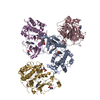


 Spodoptera frugiperda (ツマジロクサヨトウ)
 分子量: 374.439 Da / 分子数: 4 / 由来タイプ: 合成 / 式: C21H22N6O
 試料調製
試料調製 / ビームライン: MX1 / 波長: 0.9537 Å
 解析
解析