 Movie
Movie Controller

[English] 日本語
 Yorodumi
- PDB-6hmd: STRUCTURE OF PROTEIN KINASE CK2 CATALYTIC SUBUNIT (ISOFORM CK2ALP... -
+ Open data

- Basic information
| Entry | Database: PDB / ID: 6hmd | ||||||
|---|---|---|---|---|---|---|---|
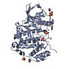
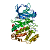
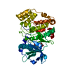
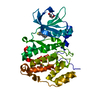
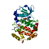
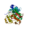
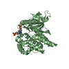
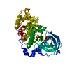
| Title | STRUCTURE OF PROTEIN KINASE CK2 CATALYTIC SUBUNIT (ISOFORM CK2ALPHA'; CSNK2A2 gene product) IN COMPLEX WITH THE INDENOINDOLE-TYPE INHIBITOR AR18 | ||||||
 Components Components | Casein kinase II subunit alpha' | ||||||
 Keywords Keywords | TRANSFERASE / protein kinase CK2 / casein kinase 2 / catalytic subunit ck2alpha' / csnk2a2 / indeno[1 / 2-b]indole-type inhibitor | ||||||
| Function / homology |  Function and homology information Function and homology informationSPOP-mediated proteasomal degradation of PD-L1(CD274) / regulation of mitophagy / regulation of chromosome separation / WNT mediated activation of DVL / Condensation of Prometaphase Chromosomes / protein kinase CK2 complex / : / Phosphorylation and nuclear translocation of the CRY:PER:kinase complex / Regulation of CDH1 posttranslational processing and trafficking to plasma membrane / Receptor Mediated Mitophagy ...SPOP-mediated proteasomal degradation of PD-L1(CD274) / regulation of mitophagy / regulation of chromosome separation / WNT mediated activation of DVL / Condensation of Prometaphase Chromosomes / protein kinase CK2 complex / : / Phosphorylation and nuclear translocation of the CRY:PER:kinase complex / Regulation of CDH1 posttranslational processing and trafficking to plasma membrane / Receptor Mediated Mitophagy / Synthesis of PC / RUNX1 interacts with co-factors whose precise effect on RUNX1 targets is not known / Maturation of hRSV A proteins / negative regulation of apoptotic signaling pathway / negative regulation of proteasomal ubiquitin-dependent protein catabolic process / liver regeneration / acrosomal vesicle / Signal transduction by L1 / cerebral cortex development / Wnt signaling pathway / Regulation of PTEN stability and activity / KEAP1-NFE2L2 pathway / double-strand break repair / Cooperation of PDCL (PhLP1) and TRiC/CCT in G-protein beta folding / heterochromatin formation / spermatogenesis / Regulation of TP53 Activity through Phosphorylation / non-specific serine/threonine protein kinase / protein serine kinase activity / protein serine/threonine kinase activity / apoptotic process / DNA damage response / positive regulation of DNA-templated transcription / chromatin / DNA-templated transcription / nucleoplasm / ATP binding / nucleus / cytosol Similarity search - Function | ||||||
| Biological species |  Homo sapiens (human) Homo sapiens (human) | ||||||
| Method |  X-RAY DIFFRACTION / SYNCHROTRON / AB INITIO PHASING / Resolution: 1 Å X-RAY DIFFRACTION / SYNCHROTRON / AB INITIO PHASING / Resolution: 1 Å | ||||||
 Authors Authors | Niefind, K. / Lindenblatt, D. / Jose, J. / Le Borgne, M. | ||||||
| Funding support |  Germany, 1items Germany, 1items
| ||||||
 Citation Citation | Journal: Acs Omega / Year: 2019 Title: Diacritic Binding of an Indenoindole Inhibitor by CK2 alpha Paralogs Explored by a Reliable Path to Atomic Resolution CK2 alpha ' Structures. Authors: Lindenblatt, D. / Nickelsen, A. / Applegate, V.M. / Hochscherf, J. / Witulski, B. / Bouaziz, Z. / Marminon, C. / Bretner, M. / Le Borgne, M. / Jose, J. / Niefind, K. #1: Journal: Pharmaceuticals (Basel) / Year: 2017Title: Unexpected Binding Mode of a Potent Indeno[1,2-b]indole-Type Inhibitor of Protein Kinase CK2 Revealed by Complex Structures with the Catalytic Subunit CK2alpha and Its Paralog CK2alpha' Authors: Hochscherf, J. / Lindenblatt, D. / Witulski, B. / Birus, R. / Aichele, D. / Marminon, C. / Bouaziz, Z. / Le Borgne, M. / Jose, J. / Niefind, K. #2: Journal: J. Mol. Biol. / Year: 2003Title: Crystal structure of a C-terminal deletion mutant of human protein kinase CK2 catalytic subunit. Authors: Ermakova, I. / Boldyreff, B. / Issinger, O.G. / Niefind, K. | ||||||
| History |
|
- Structure visualization
| Structure viewer | Molecule: MolmilJmol/JSmol |
|---|
- Downloads & links
-Download
| PDBx/mmCIF format | 6hmd.cif.gz | 246.9 KB | Display | PDBx/mmCIF format |
|---|---|---|---|---|
| PDB format | pdb6hmd.ent.gz | 199.8 KB | Display | PDB format |
| PDBx/mmJSON format | 6hmd.json.gz | Tree view | PDBx/mmJSON format | |
| Others |  Other downloads Other downloads |
-Validation report
| Arichive directory | https://data.pdbj.org/pub/pdb/validation_reports/hm/6hmdftp://data.pdbj.org/pub/pdb/validation_reports/hm/6hmd | HTTPS FTP |
|---|
-Related structure data


-Links
 PDBj
PDBj


- Assembly
| Deposited unit | 
| ||||||||
|---|---|---|---|---|---|---|---|---|---|
| 1 |
| ||||||||
| Unit cell |
|
-Components
| #1: Protein | Mass: 43018.012 Da / Num. of mol.: 1 / Mutation: C336S Source method: isolated from a genetically manipulated source Source: (gene. exp.) Homo sapiens (human) / Gene: CSNK2A2, CK2A2 / Production host:  References: UniProt: P19784, non-specific serine/threonine protein kinase | ||||||
|---|---|---|---|---|---|---|---|
| #2: Chemical |   Mass: 62.068 Da / Num. of mol.: 2 / Source method: obtained synthetically / Formula: C2H6O2 Mass: 62.068 Da / Num. of mol.: 2 / Source method: obtained synthetically / Formula: C2H6O2#3: Chemical | ChemComp-GDW / |   Mass: 336.427 Da / Num. of mol.: 1 / Source method: obtained synthetically / Formula: C21H24N2O2 Mass: 336.427 Da / Num. of mol.: 1 / Source method: obtained synthetically / Formula: C21H24N2O2#4: Chemical | ChemComp-CL / |   Mass: 35.453 Da / Num. of mol.: 1 / Source method: isolated from a natural source / Formula: Cl Mass: 35.453 Da / Num. of mol.: 1 / Source method: isolated from a natural source / Formula: Cl#5: Water | ChemComp-HOH / |  Mass: 18.015 Da / Num. of mol.: 482 / Source method: isolated from a natural source / Formula: H2O Mass: 18.015 Da / Num. of mol.: 482 / Source method: isolated from a natural source / Formula: H2O |
-Experimental details
-Experiment
| Experiment | Method: X-RAY DIFFRACTION / Number of used crystals: 1 |
|---|
- Sample preparation
| Crystal | Density Matthews: 2.24 Å3/Da / Density % sol: 44.98 % |
|---|---|
| Crystal grow | Temperature: 293.15 K / Method: vapor diffusion, sitting drop / pH: 8.5 Details: 180 MICROLITER ENZYME STOCK SOLUTION (6 MG/ML IN 500 MM NACL, 25 MM TRIS/HCL, PH 8.5) WAS MIXED and incubated WITH 20 MIKROLITER of a STOCK SOLUTION of the inhibitor 4B0 (10 MM 4B0 IN DMSO). ...Details: 180 MICROLITER ENZYME STOCK SOLUTION (6 MG/ML IN 500 MM NACL, 25 MM TRIS/HCL, PH 8.5) WAS MIXED and incubated WITH 20 MIKROLITER of a STOCK SOLUTION of the inhibitor 4B0 (10 MM 4B0 IN DMSO). 20 MICROLITERS OF the resulting solution WAS MIXED WITH 10 MICROLITERS OF RESERVOIR SOLUTION (700 MM LICL, 28 % (W/V) PEG 6000, 100 MM TRIS/HCL, PH 8.5) IN EACH WELL OF A CRYSTALLIZATION PLATE. A SINGLE MACROSEED, grown UNDER THE SAME CONDITIONS, WAS ADDED TO EACH DROPLET of the plate. THE DROPLETS WERE EQUILIBRATED AT 293.15 K using the sitting drop variant of the VAPOR DIFFUSION method. This procedure generated large single crystals of a CK2alpha'/4B0 complex. Afterwards, the inhibitor 4B0 was exchanged against AR18 by an extensive soaking procedure of several steps. |
-Data collection
| Diffraction | Mean temperature: 100 K |
|---|---|
| Diffraction source | Source: SYNCHROTRON / Site: ESRF  / Beamline: BM30A / Wavelength: 0.920042 Å / Beamline: BM30A / Wavelength: 0.920042 Å |
| Detector | Type: ADSC QUANTUM 315r / Detector: CCD / Date: Nov 23, 2017 |
| Radiation | Protocol: SINGLE WAVELENGTH / Monochromatic (M) / Laue (L): M / Scattering type: x-ray |
| Radiation wavelength | Wavelength: 0.920042 Å / Relative weight: 1 |
| Reflection | Resolution: 1→44.88 Å / Num. obs: 177359 / % possible obs: 92.18 % / Redundancy: 3.9 % / Biso Wilson estimate: 10.12 Å2 / CC1/2: 0.999 / Rmerge(I) obs: 0.04708 / Rpim(I) all: 0.02787 / Rrim(I) all: 0.05476 / Rsym value: 0.04708 / Net I/σ(I): 15.67 |
| Reflection shell | Resolution: 1→1.036 Å / Redundancy: 4 % / Rmerge(I) obs: 1.028 / Mean I/σ(I) obs: 1.36 / Num. unique obs: 17032 / CC1/2: 0.557 / Rpim(I) all: 0.5973 / Rrim(I) all: 1.189 / Rsym value: 1.028 / % possible all: 88.55 |
- Processing
| Software |
| ||||||||||||||||||||||||||||||||||||||||||||||||||||||||||||||||||||||||||||||||||||||||||||||||||
|---|---|---|---|---|---|---|---|---|---|---|---|---|---|---|---|---|---|---|---|---|---|---|---|---|---|---|---|---|---|---|---|---|---|---|---|---|---|---|---|---|---|---|---|---|---|---|---|---|---|---|---|---|---|---|---|---|---|---|---|---|---|---|---|---|---|---|---|---|---|---|---|---|---|---|---|---|---|---|---|---|---|---|---|---|---|---|---|---|---|---|---|---|---|---|---|---|---|---|---|
| Refinement | Method to determine structure: AB INITIO PHASING / Resolution: 1→22.327 Å / SU ML: 0.1 / Cross valid method: FREE R-VALUE / σ(F): 1.96 / Phase error: 18.05
| ||||||||||||||||||||||||||||||||||||||||||||||||||||||||||||||||||||||||||||||||||||||||||||||||||
| Solvent computation | Shrinkage radii: 0.9 Å / VDW probe radii: 1.11 Å | ||||||||||||||||||||||||||||||||||||||||||||||||||||||||||||||||||||||||||||||||||||||||||||||||||
| Refinement step | Cycle: LAST / Resolution: 1→22.327 Å
| ||||||||||||||||||||||||||||||||||||||||||||||||||||||||||||||||||||||||||||||||||||||||||||||||||
| Refine LS restraints |
| ||||||||||||||||||||||||||||||||||||||||||||||||||||||||||||||||||||||||||||||||||||||||||||||||||
| LS refinement shell |
|
