 Movie
Movie Controller

[English] 日本語
 Yorodumi
- PDB-6drx: Structural Determinants of Activation and Biased Agonism at the 5... -
+ Open data

- Basic information
| Entry | Database: PDB / ID: 6drx | |||||||||||||||||||||
|---|---|---|---|---|---|---|---|---|---|---|---|---|---|---|---|---|---|---|---|---|---|---|
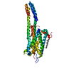
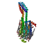
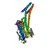
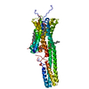
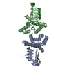
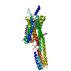
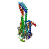
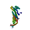
| Title | Structural Determinants of Activation and Biased Agonism at the 5-HT2B Receptor | |||||||||||||||||||||
 Components Components | 5HT2B receptor, BRIL chimera | |||||||||||||||||||||
 Keywords Keywords | MEMBRANE PROTEIN / GPCR / 5HT2B / Setotonin receptor / Lisuride | |||||||||||||||||||||
| Function / homology |  Function and homology information Function and homology informationintestine smooth muscle contraction / Gq/11-coupled serotonin receptor activity / positive regulation of phosphatidylinositol biosynthetic process / G protein-coupled serotonin receptor signaling pathway / G protein-coupled serotonin receptor complex / regulation of behavior / neural crest cell differentiation / phospholipase C-activating serotonin receptor signaling pathway / embryonic morphogenesis / serotonin receptor activity ...intestine smooth muscle contraction / Gq/11-coupled serotonin receptor activity / positive regulation of phosphatidylinositol biosynthetic process / G protein-coupled serotonin receptor signaling pathway / G protein-coupled serotonin receptor complex / regulation of behavior / neural crest cell differentiation / phospholipase C-activating serotonin receptor signaling pathway / embryonic morphogenesis / serotonin receptor activity / Serotonin receptors / G protein-coupled serotonin receptor activity / vasoconstriction / serotonin receptor signaling pathway / neural crest cell migration / neurotransmitter receptor activity / cardiac muscle hypertrophy / serotonin binding / positive regulation of cell division / heart morphogenesis / ERK1 and ERK2 cascade / G-protein alpha-subunit binding / G protein-coupled receptor signaling pathway, coupled to cyclic nucleotide second messenger / release of sequestered calcium ion into cytosol / positive regulation of endothelial cell proliferation / positive regulation of cytokine production / GTPase activator activity / calcium-mediated signaling / electron transport chain / intracellular calcium ion homeostasis / chemical synaptic transmission / G alpha (q) signalling events / positive regulation of ERK1 and ERK2 cascade / positive regulation of canonical NF-kappaB signal transduction / electron transfer activity / periplasmic space / response to xenobiotic stimulus / iron ion binding / G protein-coupled receptor signaling pathway / heme binding / positive regulation of cell population proliferation / synapse / dendrite / negative regulation of apoptotic process / nucleoplasm / plasma membrane / cytoplasm Similarity search - Function | |||||||||||||||||||||
| Biological species |  Homo sapiens (human) Homo sapiens (human) | |||||||||||||||||||||
| Method |  X-RAY DIFFRACTION / SYNCHROTRON / MOLECULAR REPLACEMENT / Resolution: 3.1 Å X-RAY DIFFRACTION / SYNCHROTRON / MOLECULAR REPLACEMENT / Resolution: 3.1 Å | |||||||||||||||||||||
 Authors Authors | McCorvy, J.D. / Wacker, D. / Wang, S. / Agegnehu, B. / Liu, J. / Lansu, K. / Tribo, A.R. / Olsen, R.H.J. / Che, T. / Jin, J. / Roth, B.L. | |||||||||||||||||||||
| Funding support |  United States, 6items United States, 6items
| |||||||||||||||||||||
 Citation Citation | Journal: Nat. Struct. Mol. Biol. / Year: 2018 Title: Structural determinants of 5-HT2Breceptor activation and biased agonism. Authors: McCorvy, J.D. / Wacker, D. / Wang, S. / Agegnehu, B. / Liu, J. / Lansu, K. / Tribo, A.R. / Olsen, R.H.J. / Che, T. / Jin, J. / Roth, B.L. | |||||||||||||||||||||
| History |
|
- Structure visualization
| Structure viewer | Molecule: MolmilJmol/JSmol |
|---|
- Downloads & links
-Download
| PDBx/mmCIF format | 6drx.cif.gz | 156.1 KB | Display | PDBx/mmCIF format |
|---|---|---|---|---|
| PDB format | pdb6drx.ent.gz | 119.6 KB | Display | PDB format |
| PDBx/mmJSON format | 6drx.json.gz | Tree view | PDBx/mmJSON format | |
| Others |  Other downloads Other downloads |
-Validation report
| Arichive directory | https://data.pdbj.org/pub/pdb/validation_reports/dr/6drxftp://data.pdbj.org/pub/pdb/validation_reports/dr/6drx | HTTPS FTP |
|---|
-Related structure data
| Related structure data |  6dryC  6drzC  6ds0C 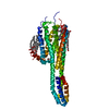 4ib4S S: Starting model for refinement C: citing same article ( |
|---|---|
| Similar structure data |
-Links
 PDBj
PDBj


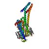
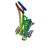
- Assembly
| Deposited unit | 
| ||||||||
|---|---|---|---|---|---|---|---|---|---|
| 1 |
| ||||||||
| Unit cell |
|
-Components
| #1: Protein | Mass: 46049.887 Da / Num. of mol.: 1 Source method: isolated from a genetically manipulated source Source: (gene. exp.) Homo sapiens (human), (gene. exp.) Gene: HTR2B, cybC / Production host:   Spodoptera frugiperda (fall armyworm) / References: UniProt: P41595, UniProt: P0ABE7 Spodoptera frugiperda (fall armyworm) / References: UniProt: P41595, UniProt: P0ABE7 |
|---|---|
| #2: Chemical | ChemComp-H8G /   Mass: 338.447 Da / Num. of mol.: 1 / Source method: obtained synthetically / Formula: C20H26N4O / Comment: medication*YM Mass: 338.447 Da / Num. of mol.: 1 / Source method: obtained synthetically / Formula: C20H26N4O / Comment: medication*YM |
| #3: Chemical | ChemComp-CLR /   Mass: 386.654 Da / Num. of mol.: 1 / Source method: obtained synthetically / Formula: C27H46O Mass: 386.654 Da / Num. of mol.: 1 / Source method: obtained synthetically / Formula: C27H46O |
| #4: Chemical | ChemComp-OLC / (  Mass: 356.540 Da / Num. of mol.: 1 / Source method: obtained synthetically / Formula: C21H40O4 Mass: 356.540 Da / Num. of mol.: 1 / Source method: obtained synthetically / Formula: C21H40O4 |
| #5: Water | ChemComp-HOH /  Mass: 18.015 Da / Num. of mol.: 1 / Source method: isolated from a natural source / Formula: H2O Mass: 18.015 Da / Num. of mol.: 1 / Source method: isolated from a natural source / Formula: H2O |
| Has protein modification | Y |
-Experimental details
-Experiment
| Experiment | Method: X-RAY DIFFRACTION / Number of used crystals: 1 |
|---|
- Sample preparation
| Crystal | Density Matthews: 3.21 Å3/Da / Density % sol: 61.73 % |
|---|---|
| Crystal grow | Temperature: 293 K / Method: lipidic cubic phase Details: 100 mM Tris/HCl pH 7.4-7.7, 30-50 mM Ammonium tartrate dibasic, 30% v/v PEG400 |
-Data collection
| Diffraction | Mean temperature: 100 K |
|---|---|
| Diffraction source | Source: SYNCHROTRON / Site: APS / Beamline: 23-ID-B / Wavelength: 1.033 Å |
| Detector | Type: DECTRIS EIGER X 16M / Detector: PIXEL / Date: Aug 4, 2016 |
| Radiation | Protocol: SINGLE WAVELENGTH / Monochromatic (M) / Laue (L): M / Scattering type: x-ray |
| Radiation wavelength | Wavelength: 1.033 Å / Relative weight: 1 |
| Reflection | Resolution: 3.1→30 Å / Num. obs: 10701 / % possible obs: 97.1 % / Redundancy: 4.4 % / CC1/2: 0.996 / Rmerge(I) obs: 0.122 / Net I/σ(I): 10.9 |
| Reflection shell | Resolution: 3.1→3.17 Å / Redundancy: 4.3 % / Rmerge(I) obs: 0.95 / Mean I/σ(I) obs: 1.1 / Num. unique obs: 699 / CC1/2: 0.462 / % possible all: 97.1 |
- Processing
| Software |
| ||||||||||||||||||||||||||||||||||||||||||||||||||||||||||||||||||||||||||||||||||||||||||||||||||||
|---|---|---|---|---|---|---|---|---|---|---|---|---|---|---|---|---|---|---|---|---|---|---|---|---|---|---|---|---|---|---|---|---|---|---|---|---|---|---|---|---|---|---|---|---|---|---|---|---|---|---|---|---|---|---|---|---|---|---|---|---|---|---|---|---|---|---|---|---|---|---|---|---|---|---|---|---|---|---|---|---|---|---|---|---|---|---|---|---|---|---|---|---|---|---|---|---|---|---|---|---|---|
| Refinement | Method to determine structure: MOLECULAR REPLACEMENT Starting model: PDB entry: 4IB4 Resolution: 3.1→29.257 Å / SU ML: 0.5 / Cross valid method: FREE R-VALUE / σ(F): 1.37 / Phase error: 28.99
| ||||||||||||||||||||||||||||||||||||||||||||||||||||||||||||||||||||||||||||||||||||||||||||||||||||
| Solvent computation | Shrinkage radii: 0.9 Å / VDW probe radii: 1.11 Å | ||||||||||||||||||||||||||||||||||||||||||||||||||||||||||||||||||||||||||||||||||||||||||||||||||||
| Refinement step | Cycle: LAST / Resolution: 3.1→29.257 Å
| ||||||||||||||||||||||||||||||||||||||||||||||||||||||||||||||||||||||||||||||||||||||||||||||||||||
| Refine LS restraints |
| ||||||||||||||||||||||||||||||||||||||||||||||||||||||||||||||||||||||||||||||||||||||||||||||||||||
| LS refinement shell |
| ||||||||||||||||||||||||||||||||||||||||||||||||||||||||||||||||||||||||||||||||||||||||||||||||||||
| Refinement TLS params. | Method: refined / Refine-ID: X-RAY DIFFRACTION
| ||||||||||||||||||||||||||||||||||||||||||||||||||||||||||||||||||||||||||||||||||||||||||||||||||||
| Refinement TLS group |
|
