ムービー
ムービー コントローラー


+ データを開く

- 基本情報
基本情報
| 登録情報 |  | |||||||||
|---|---|---|---|---|---|---|---|---|---|---|
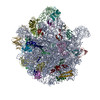
| タイトル | Xanthomonas citri pv. glycines 50S subunit | |||||||||
 マップデータ マップデータ | Xanthomonas campestris 50S map | |||||||||
 試料 試料 |
| |||||||||
 キーワード キーワード | Phytopathogen / Xanthomonas citri pv. glycines / 50S subunit / RIBOSOME | |||||||||
| 機能・相同性 |  機能・相同性情報 機能・相同性情報: / : / : / : / : / : / : / : / : / : ...: / : / : / : / : / : / : / : / : / : / : / : / : / : / : / : / : / : / : / : / : / : / : / : / : / : / : 類似検索 - 構成要素 | |||||||||
| 生物種 |  Xanthomonas citri pv. glycines (バクテリア) Xanthomonas citri pv. glycines (バクテリア) | |||||||||
| 手法 | 単粒子再構成法 / クライオ電子顕微鏡法 / 解像度: 3.0 Å | |||||||||
 データ登録者 データ登録者 | Sengupta S / Anand R | |||||||||
| 資金援助 |  インド, 1件 インド, 1件
| |||||||||
 引用 引用 | ジャーナル: Acs Omega / 年: 2026 タイトル: Structure of 50S ribosomal subunit from phytopathogen Xanthomonas citri pv. glycines 著者: Sengupta S / Tripathy J / Bandyopadhay N / Gautam S / Anand R | |||||||||
| 履歴 |
|
- 構造の表示
構造の表示
| 添付画像 |
|---|

- ダウンロードとリンク
-EMDBアーカイブ
| マップデータ | emd_69918.map.gz | 27.9 MB | EMDBマップデータ形式 | |
|---|---|---|---|---|
| ヘッダ (付随情報) | emd-69918-v30.xmlemd-69918.xml | 45.2 KB 45.2 KB | 表示 表示 | EMDBヘッダ |
| FSC (解像度算出) | emd_69918_fsc.xml | 14.1 KB | 表示 | FSCデータファイル |
| 画像 | 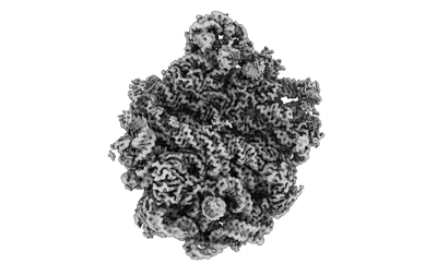 emd_69918.png emd_69918.png | 61.8 KB | ||
| Filedesc metadata | emd-69918.cif.gz | 9.8 KB | ||
| その他 | emd_69918_half_map_1.map.gzemd_69918_half_map_2.map.gz | 194 MB 194 MB | ||
| アーカイブディレクトリ |  https://data.pdbj.org/pub/emdb/structures/EMD-69918ftp://data.pdbj.org/pub/emdb/structures/EMD-69918 https://data.pdbj.org/pub/emdb/structures/EMD-69918ftp://data.pdbj.org/pub/emdb/structures/EMD-69918 | HTTPS FTP |
-関連構造データ

-リンク
| EMDBのページ | EMDB (EBI/PDBe) / EMDataResource |
|---|
-マップ
| ファイル | ダウンロード / ファイル: emd_69918.map.gz / 形式: CCP4 / 大きさ: 244.1 MB / タイプ: IMAGE STORED AS FLOATING POINT NUMBER (4 BYTES) | ||||||||||||||||||||||||||||||||||||
|---|---|---|---|---|---|---|---|---|---|---|---|---|---|---|---|---|---|---|---|---|---|---|---|---|---|---|---|---|---|---|---|---|---|---|---|---|---|
| 注釈 | Xanthomonas campestris 50S map | ||||||||||||||||||||||||||||||||||||
| 投影像・断面図 | 画像のコントロール
画像は Spider により作成 | ||||||||||||||||||||||||||||||||||||
| ボクセルのサイズ | X=Y=Z: 0.85 Å | ||||||||||||||||||||||||||||||||||||
| 密度 |
| ||||||||||||||||||||||||||||||||||||
| 対称性 | 空間群: 1 | ||||||||||||||||||||||||||||||||||||
| 詳細 | EMDB XML:
|
 Z (Sec.)
 Y (Row.)
 X (Col.)


-添付データ
-ハーフマップ: #2
| ファイル | emd_69918_half_map_1.map | ||||||||||||
|---|---|---|---|---|---|---|---|---|---|---|---|---|---|
| 投影像・断面図 |
| ||||||||||||
| 密度ヒストグラム |


-ハーフマップ: #1
| ファイル | emd_69918_half_map_2.map | ||||||||||||
|---|---|---|---|---|---|---|---|---|---|---|---|---|---|
| 投影像・断面図 |
| ||||||||||||
| 密度ヒストグラム |


- 試料の構成要素
+全体 : 50S rebosomal subunit of Xanthomonas citri pv. glycine
+超分子 #1: 50S rebosomal subunit of Xanthomonas citri pv. glycine
+分子 #1: Large ribosomal subunit protein bL32
+分子 #2: Large ribosomal subunit protein bL33
+分子 #3: Large ribosomal subunit protein bL34
+分子 #4: Large ribosomal subunit protein bL35
+分子 #5: Large ribosomal subunit protein bL36
+分子 #8: Large ribosomal subunit protein uL2
+分子 #9: Large ribosomal subunit protein uL3
+分子 #10: Large ribosomal subunit protein uL4
+分子 #11: Large ribosomal subunit protein uL6
+分子 #12: Large ribosomal subunit protein bL9
+分子 #13: Large ribosomal subunit protein uL13
+分子 #14: Large ribosomal subunit protein uL14
+分子 #15: Large ribosomal subunit protein uL15
+分子 #16: Large ribosomal subunit protein uL16
+分子 #17: Large ribosomal subunit protein bL17
+分子 #18: Large ribosomal subunit protein uL18
+分子 #19: Large ribosomal subunit protein bL19
+分子 #20: Large ribosomal subunit protein bL20
+分子 #21: Large ribosomal subunit protein bL21
+分子 #22: Large ribosomal subunit protein uL22
+分子 #23: Large ribosomal subunit protein uL23
+分子 #24: Large ribosomal subunit protein uL24
+分子 #25: Large ribosomal subunit protein bL25
+分子 #26: Large ribosomal subunit protein bL27
+分子 #27: Large ribosomal subunit protein bL28
+分子 #28: Large ribosomal subunit protein uL29
+分子 #29: Large ribosomal subunit protein uL30
+分子 #6: RNA (2797-MER)
+分子 #7: RNA (117-MER)
-実験情報
-構造解析
| 手法 | クライオ電子顕微鏡法 |
|---|---|
 解析 解析 | 単粒子再構成法 |
| 試料の集合状態 | particle |
-試料調製
| 緩衝液 | pH: 7.5 |
|---|---|
| 凍結 | 凍結剤: ETHANE |
- 電子顕微鏡法
| 顕微鏡 | TFS KRIOS |
|---|---|
| 特殊光学系 | エネルギーフィルター - 名称: GIF Bioquantum |
| 撮影 | フィルム・検出器のモデル: GATAN K3 BIOCONTINUUM (6k x 4k) 平均電子線量: 43.0 e/Å2 |
| 電子線 | 加速電圧: 300 kV / 電子線源:  FIELD EMISSION GUN FIELD EMISSION GUN |
| 電子光学系 | 照射モード: FLOOD BEAM / 撮影モード: BRIGHT FIELD / 最大 デフォーカス(公称値): 2.0 µm / 最小 デフォーカス(公称値): 0.5 µm |
| 実験機器 |  モデル: Titan Krios / 画像提供: FEI Company |