ムービー
ムービー コントローラー


+ データを開く

- 基本情報
基本情報
| 登録情報 | データベース: EMDB / ID: EMD-6526 | |||||||||
|---|---|---|---|---|---|---|---|---|---|---|
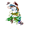
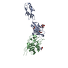
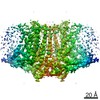
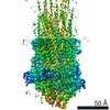
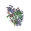
| タイトル | Cryo-electron microscopy structure of a coronavirus spike glycoprotein trimer | |||||||||
 マップデータ マップデータ | Reconstruction of a furin cleavage site mutant | |||||||||
 試料 試料 |
| |||||||||
 キーワード キーワード | Coronavirus / viral fusion proteins / viral spike / peplomer | |||||||||
| 機能・相同性 |  機能・相同性情報 機能・相同性情報endocytosis involved in viral entry into host cell / host cell Golgi apparatus / host cell endoplasmic reticulum-Golgi intermediate compartment membrane / receptor-mediated virion attachment to host cell / fusion of virus membrane with host plasma membrane / fusion of virus membrane with host endosome membrane / viral envelope / host cell plasma membrane / virion membrane / identical protein binding / membrane 類似検索 - 分子機能 | |||||||||
| 生物種 |  Murine hepatitis virus (マウス肝炎ウイルス) Murine hepatitis virus (マウス肝炎ウイルス) | |||||||||
| 手法 | 単粒子再構成法 / クライオ電子顕微鏡法 / 解像度: 4.0 Å | |||||||||
 データ登録者 データ登録者 | Walls AC / Tortorici MA / Bosch BJ / Frenz BJ / Rottier PJM / DiMaio F / Rey FA / Veesler D | |||||||||
 引用 引用 | ジャーナル: Nature / 年: 2016 タイトル: Cryo-electron microscopy structure of a coronavirus spike glycoprotein trimer. 著者: Alexandra C Walls / M Alejandra Tortorici / Berend-Jan Bosch / Brandon Frenz / Peter J M Rottier / Frank DiMaio / Félix A Rey / David Veesler /    要旨: The tremendous pandemic potential of coronaviruses was demonstrated twice in the past few decades by two global outbreaks of deadly pneumonia. Entry of coronaviruses into cells is mediated by the ...The tremendous pandemic potential of coronaviruses was demonstrated twice in the past few decades by two global outbreaks of deadly pneumonia. Entry of coronaviruses into cells is mediated by the transmembrane spike glycoprotein S, which forms a trimer carrying receptor-binding and membrane fusion functions. S also contains the principal antigenic determinants and is the target of neutralizing antibodies. Here we present the structure of a mouse coronavirus S trimer ectodomain determined at 4.0 Å resolution by single particle cryo-electron microscopy. It reveals the metastable pre-fusion architecture of S and highlights key interactions stabilizing it. The structure shares a common core with paramyxovirus F proteins, implicating mechanistic similarities and an evolutionary connection between these viral fusion proteins. The accessibility of the highly conserved fusion peptide at the periphery of the trimer indicates potential vaccinology strategies to elicit broadly neutralizing antibodies against coronaviruses. Finally, comparison with crystal structures of human coronavirus S domains allows rationalization of the molecular basis for species specificity based on the use of spatially contiguous but distinct domains. | |||||||||
| 履歴 |
|
- 構造の表示
構造の表示
| ムービー |
ムービービューア |
|---|---|
| 構造ビューア | EMマップ: SurfViewMolmilJmol/JSmol |
| 添付画像 |

- ダウンロードとリンク
-EMDBアーカイブ
| マップデータ | emd_6526.map.gz | 8.3 MB | EMDBマップデータ形式 | |
|---|---|---|---|---|
| ヘッダ (付随情報) | emd-6526-v30.xmlemd-6526.xml | 11 KB 11 KB | 表示 表示 | EMDBヘッダ |
| 画像 |  400_6526.gif 400_6526.gif 80_6526.gif 80_6526.gif | 81.9 KB 4.9 KB | ||
| アーカイブディレクトリ |  http://ftp.pdbj.org/pub/emdb/structures/EMD-6526ftp://ftp.pdbj.org/pub/emdb/structures/EMD-6526 http://ftp.pdbj.org/pub/emdb/structures/EMD-6526ftp://ftp.pdbj.org/pub/emdb/structures/EMD-6526 | HTTPS FTP |
-関連構造データ
-リンク
| EMDBのページ | EMDB (EBI/PDBe) / EMDataResource |
|---|
-マップ
| ファイル | ダウンロード / ファイル: emd_6526.map.gz / 形式: CCP4 / 大きさ: 89 MB / タイプ: IMAGE STORED AS FLOATING POINT NUMBER (4 BYTES) | ||||||||||||||||||||||||||||||||||||||||||||||||||||||||||||||||||||
|---|---|---|---|---|---|---|---|---|---|---|---|---|---|---|---|---|---|---|---|---|---|---|---|---|---|---|---|---|---|---|---|---|---|---|---|---|---|---|---|---|---|---|---|---|---|---|---|---|---|---|---|---|---|---|---|---|---|---|---|---|---|---|---|---|---|---|---|---|---|
| 注釈 | Reconstruction of a furin cleavage site mutant | ||||||||||||||||||||||||||||||||||||||||||||||||||||||||||||||||||||
| ボクセルのサイズ | X=Y=Z: 1.46 Å | ||||||||||||||||||||||||||||||||||||||||||||||||||||||||||||||||||||
| 密度 |
| ||||||||||||||||||||||||||||||||||||||||||||||||||||||||||||||||||||
| 対称性 | 空間群: 1 | ||||||||||||||||||||||||||||||||||||||||||||||||||||||||||||||||||||
| 詳細 | EMDB XML:
CCP4マップ ヘッダ情報:
| ||||||||||||||||||||||||||||||||||||||||||||||||||||||||||||||||||||
-添付データ
- 試料の構成要素
-全体 : Murine hepatitis virus protein S
| 全体 | 名称: Murine hepatitis virus protein S |
|---|---|
| 要素 |
|
-超分子 #1000: Murine hepatitis virus protein S
| 超分子 | 名称: Murine hepatitis virus protein S / タイプ: sample / ID: 1000 / 集合状態: Trimer / Number unique components: 1 |
|---|---|
| 分子量 | 理論値: 420 KDa |
-分子 #1: Murine hepatitis virus protein S
| 分子 | 名称: Murine hepatitis virus protein S / タイプ: protein_or_peptide / ID: 1 / 集合状態: Trimer / 組換発現: Yes |
|---|---|
| 由来(天然) | 生物種: Murine hepatitis virus (マウス肝炎ウイルス) 株: A59 / 別称: MHV |
| 分子量 | 理論値: 140 KDa |
| 組換発現 | 生物種:  組換細胞: S2 |
| 配列 | UniProtKB: Spike glycoprotein |
-実験情報
-構造解析
| 手法 | クライオ電子顕微鏡法 |
|---|---|
 解析 解析 | 単粒子再構成法 |
| 試料の集合状態 | particle |
-試料調製
| 濃度 | 1.85 mg/mL |
|---|---|
| 緩衝液 | pH: 7.5 / 詳細: 20 mM Tris-HCl, 100 mM NaCl |
| 凍結 | 凍結剤: ETHANE / チャンバー内湿度: 90 % / チャンバー内温度: 93 K / 装置: GATAN CRYOPLUNGE 3 / 手法: Blot for 3.5 seconds before plunging |
- 電子顕微鏡法
| 顕微鏡 | FEI TITAN KRIOS |
|---|---|
| 日付 | 2014年12月9日 |
| 撮影 | カテゴリ: CCD フィルム・検出器のモデル: GATAN K2 SUMMIT (4k x 4k) 実像数: 1600 / 平均電子線量: 53 e/Å2 詳細: Every image is the average of 38 frames recorded by the direct electron detector. |
| 電子線 | 加速電圧: 300 kV / 電子線源:  FIELD EMISSION GUN FIELD EMISSION GUN |
| 電子光学系 | 倍率(補正後): 38022 / 照射モード: FLOOD BEAM / 撮影モード: BRIGHT FIELD / Cs: 2.7 mm / 最大 デフォーカス(公称値): 5.0 µm / 最小 デフォーカス(公称値): 2.0 µm / 倍率(公称値): 22500 |
| 試料ステージ | 試料ホルダーモデル: FEI TITAN KRIOS AUTOGRID HOLDER |
| 実験機器 |  モデル: Titan Krios / 画像提供: FEI Company |
-画像解析
| CTF補正 | 詳細: Each particle |
|---|---|
| 最終 再構成 | アルゴリズム: OTHER / 解像度のタイプ: BY AUTHOR / 解像度: 4.0 Å / 解像度の算出法: OTHER / ソフトウェア - 名称: Relion / 使用した粒子像数: 82000 |
-原子モデル構築 1
| 初期モデル | PDB ID: |
|---|---|
| ソフトウェア | 名称: Rosetta, Coot, Chimera |
| 精密化 | 空間: REAL / プロトコル: FLEXIBLE FIT |
| 得られたモデル | 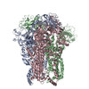 PDB-3jcl: |
-原子モデル構築 2
| 初期モデル | PDB ID: |
|---|---|
| ソフトウェア | 名称: Rosetta, Coot, Chimera |
| 精密化 | 空間: REAL / プロトコル: FLEXIBLE FIT |
| 得られたモデル | PDB-3jcl: |