 Movie
Movie Controller


+ Open data

- Basic information
| Entry | Database: PDB / ID: 5zgb | ||||||||||||||||||||||||||||||||||||||||||||||||||||||
|---|---|---|---|---|---|---|---|---|---|---|---|---|---|---|---|---|---|---|---|---|---|---|---|---|---|---|---|---|---|---|---|---|---|---|---|---|---|---|---|---|---|---|---|---|---|---|---|---|---|---|---|---|---|---|---|
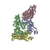
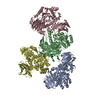
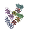
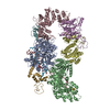
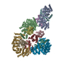
| Title | Cryo-EM structure of the red algal PSI-LHCR | ||||||||||||||||||||||||||||||||||||||||||||||||||||||
 Components Components |
| ||||||||||||||||||||||||||||||||||||||||||||||||||||||
 Keywords Keywords | PHOTOSYNTHESIS / super-complex / red alga / PSI-5Lhcr | ||||||||||||||||||||||||||||||||||||||||||||||||||||||
| Function / homology |  Function and homology information Function and homology informationplastid thylakoid membrane / thylakoid membrane / photosynthesis, light harvesting / photosystem I reaction center / photosystem I / photosynthetic electron transport in photosystem I / photosystem I / chlorophyll binding / chloroplast thylakoid membrane / photosynthesis ...plastid thylakoid membrane / thylakoid membrane / photosynthesis, light harvesting / photosystem I reaction center / photosystem I / photosynthetic electron transport in photosystem I / photosystem I / chlorophyll binding / chloroplast thylakoid membrane / photosynthesis / chloroplast / 4 iron, 4 sulfur cluster binding / electron transfer activity / oxidoreductase activity / magnesium ion binding / membrane / metal ion binding Similarity search - Function | ||||||||||||||||||||||||||||||||||||||||||||||||||||||
| Biological species |  Cyanidioschyzon merolae (eukaryote)Cyanidioschyzon merolae strain 10D (eukaryote) Cyanidioschyzon merolae (eukaryote)Cyanidioschyzon merolae strain 10D (eukaryote) | ||||||||||||||||||||||||||||||||||||||||||||||||||||||
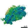
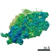
| Method | ELECTRON MICROSCOPY / single particle reconstruction / cryo EM / Resolution: 3.63 Å | ||||||||||||||||||||||||||||||||||||||||||||||||||||||
 Authors Authors | Pi, X. | ||||||||||||||||||||||||||||||||||||||||||||||||||||||
 Citation Citation | Journal: Proc Natl Acad Sci U S A / Year: 2018 Title: Unique organization of photosystem I-light-harvesting supercomplex revealed by cryo-EM from a red alga. Authors: Xiong Pi / Lirong Tian / Huai-En Dai / Xiaochun Qin / Lingpeng Cheng / Tingyun Kuang / Sen-Fang Sui / Jian-Ren Shen /   Abstract: Photosystem I (PSI) is one of the two photosystems present in oxygenic photosynthetic organisms and functions to harvest and convert light energy into chemical energy in photosynthesis. In eukaryotic ...Photosystem I (PSI) is one of the two photosystems present in oxygenic photosynthetic organisms and functions to harvest and convert light energy into chemical energy in photosynthesis. In eukaryotic algae and higher plants, PSI consists of a core surrounded by variable species and numbers of light-harvesting complex (LHC)I proteins, forming a PSI-LHCI supercomplex. Here, we report cryo-EM structures of PSI-LHCR from the red alga in two forms, one with three Lhcr subunits attached to the side, similar to that of higher plants, and the other with two additional Lhcr subunits attached to the opposite side, indicating an ancient form of PSI-LHCI. Furthermore, the red algal PSI core showed features of both cyanobacterial and higher plant PSI, suggesting an intermediate type during evolution from prokaryotes to eukaryotes. The structure of PsaO, existing in eukaryotic organisms, was identified in the PSI core and binds three chlorophylls and may be important in harvesting energy and in mediating energy transfer from LHCII to the PSI core under state-2 conditions. Individual attaching sites of LHCRs with the core subunits were identified, and each Lhcr was found to contain 11 to 13 chlorophylls and 5 zeaxanthins, which are apparently different from those of LHCs in plant PSI-LHCI. Together, our results reveal unique energy transfer pathways different from those of higher plant PSI-LHCI, its adaptation to the changing environment, and the possible changes of PSI-LHCI during evolution from prokaryotes to eukaryotes. | ||||||||||||||||||||||||||||||||||||||||||||||||||||||
| History |
|
- Structure visualization
| Movie |
Movie viewer |
|---|---|
| Structure viewer | Molecule: MolmilJmol/JSmol |
- Downloads & links
-Download
| PDBx/mmCIF format | 5zgb.cif.gz | 773.8 KB | Display | PDBx/mmCIF format |
|---|---|---|---|---|
| PDB format | pdb5zgb.ent.gz | 684.1 KB | Display | PDB format |
| PDBx/mmJSON format | 5zgb.json.gz | Tree view | PDBx/mmJSON format | |
| Others |  Other downloads Other downloads |
-Validation report
| Arichive directory | https://data.pdbj.org/pub/pdb/validation_reports/zg/5zgbftp://data.pdbj.org/pub/pdb/validation_reports/zg/5zgb | HTTPS FTP |
|---|
-Related structure data
| Related structure data | 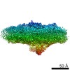 6929MC 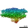 6930C 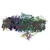 5zghC M: map data used to model this data C: citing same article ( |
|---|---|
| Similar structure data |

-Links
 PDBj
PDBj


- Assembly
| Deposited unit | 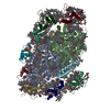
|
|---|---|
| 1 |
|
-Components
-Protein , 12 types, 14 molecules ABCDEFKLO14253
| #1: Protein | Mass: 82763.461 Da / Num. of mol.: 1 / Source method: isolated from a natural source Source: (natural) Cyanidioschyzon merolae (strain 10D) (eukaryote)Strain: 10D / References: UniProt: Q85FY7, photosystem I | ||||
|---|---|---|---|---|---|
| #2: Protein | Mass: 82107.852 Da / Num. of mol.: 1 / Source method: isolated from a natural source Source: (natural) Cyanidioschyzon merolae (strain 10D) (eukaryote)Strain: 10D / References: UniProt: Q85FY6, photosystem I | ||||
| #3: Protein | Mass: 8822.272 Da / Num. of mol.: 1 / Source method: isolated from a natural source Source: (natural) Cyanidioschyzon merolae (strain 10D) (eukaryote)Strain: 10D / References: UniProt: Q85G47, photosystem I | ||||
| #4: Protein | Mass: 15698.932 Da / Num. of mol.: 1 / Source method: isolated from a natural source Source: (natural) Cyanidioschyzon merolae (strain 10D) (eukaryote)Strain: 10D / References: UniProt: Q85FY0 | ||||
| #5: Protein | Mass: 10545.162 Da / Num. of mol.: 1 / Source method: isolated from a natural source Source: (natural) Cyanidioschyzon merolae (strain 10D) (eukaryote)Strain: 10D / References: UniProt: Q85FZ1 | ||||
| #6: Protein | Mass: 21239.318 Da / Num. of mol.: 1 / Source method: isolated from a natural source Source: (natural) Cyanidioschyzon merolae (strain 10D) (eukaryote)Strain: 10D / References: UniProt: Q85FS9 | ||||
| #9: Protein | Mass: 6295.657 Da / Num. of mol.: 1 / Source method: isolated from a natural source Source: (natural) Cyanidioschyzon merolae (strain 10D) (eukaryote)Strain: 10D / References: UniProt: Q85G51 | ||||
| #10: Protein | Mass: 15157.522 Da / Num. of mol.: 1 / Source method: isolated from a natural source Source: (natural) Cyanidioschyzon merolae (strain 10D) (eukaryote)Strain: 10D / References: UniProt: Q85FP8 | ||||
| #12: Protein | Mass: 16744.471 Da / Num. of mol.: 1 / Source method: isolated from a natural source Source: (natural) Cyanidioschyzon merolae (strain 10D) (eukaryote)Strain: 10D / References: UniProt: M1VFJ4 | ||||
| #13: Protein | Mass: 19808.818 Da / Num. of mol.: 2 / Source method: isolated from a natural source Source: (natural) Cyanidioschyzon merolae (strain 10D) (eukaryote)Strain: 10D / References: UniProt: M1VKK5 #14: Protein | Mass: 21960.398 Da / Num. of mol.: 2 / Source method: isolated from a natural source Source: (natural) Cyanidioschyzon merolae (strain 10D) (eukaryote)Strain: 10D / References: UniProt: M1UU36 #15: Protein | | Mass: 20458.535 Da / Num. of mol.: 1 / Source method: isolated from a natural source Source: (natural) Cyanidioschyzon merolae strain 10D (eukaryote)Strain: 10D |
-Protein/peptide , 3 types, 3 molecules IJM
| #7: Protein/peptide | Mass: 3408.184 Da / Num. of mol.: 1 / Source method: isolated from a natural source Source: (natural) Cyanidioschyzon merolae (strain 10D) (eukaryote)Strain: 10D / References: UniProt: Q85FQ6 |
|---|---|
| #8: Protein/peptide | Mass: 4410.245 Da / Num. of mol.: 1 / Source method: isolated from a natural source Source: (natural) Cyanidioschyzon merolae (strain 10D) (eukaryote)Strain: 10D / References: UniProt: Q85FS8 |
| #11: Protein/peptide | Mass: 3139.878 Da / Num. of mol.: 1 / Source method: isolated from a natural source Source: (natural) Cyanidioschyzon merolae (strain 10D) (eukaryote)Strain: 10D / References: UniProt: Q85G73 |
-Sugars , 2 types, 2 molecules 

| #22: Sugar | ChemComp-BGC /  Type: D-saccharide, beta linking / Mass: 180.156 Da / Num. of mol.: 1 Type: D-saccharide, beta linking / Mass: 180.156 Da / Num. of mol.: 1Source method: isolated from a genetically manipulated source Formula: C6H12O6 |
|---|---|
| #23: Sugar | ChemComp-DGD /  Type: saccharide / Mass: 949.299 Da / Num. of mol.: 1 / Source method: obtained synthetically / Formula: C51H96O15 Type: saccharide / Mass: 949.299 Da / Num. of mol.: 1 / Source method: obtained synthetically / Formula: C51H96O15 |
-Non-polymers , 9 types, 213 molecules 


| #16: Chemical | ChemComp-CL0 /  Mass: 893.489 Da / Num. of mol.: 1 / Source method: obtained synthetically / Formula: C55H72MgN4O5 Mass: 893.489 Da / Num. of mol.: 1 / Source method: obtained synthetically / Formula: C55H72MgN4O5 | ||||||||||||||
|---|---|---|---|---|---|---|---|---|---|---|---|---|---|---|---|
| #17: Chemical | ChemComp-CLA /  Mass: 893.489 Da / Num. of mol.: 157 / Source method: obtained synthetically / Formula: C55H72MgN4O5 Mass: 893.489 Da / Num. of mol.: 157 / Source method: obtained synthetically / Formula: C55H72MgN4O5#18: Chemical |  Mass: 450.696 Da / Num. of mol.: 2 / Source method: obtained synthetically / Formula: C31H46O2 Mass: 450.696 Da / Num. of mol.: 2 / Source method: obtained synthetically / Formula: C31H46O2#19: Chemical |  Mass: 722.970 Da / Num. of mol.: 2 / Source method: obtained synthetically / Formula: C38H75O10P / Comment: phospholipid*YM Mass: 722.970 Da / Num. of mol.: 2 / Source method: obtained synthetically / Formula: C38H75O10P / Comment: phospholipid*YM#20: Chemical | ChemComp-BCR /  Mass: 536.873 Da / Num. of mol.: 21 / Source method: obtained synthetically / Formula: C40H56 Mass: 536.873 Da / Num. of mol.: 21 / Source method: obtained synthetically / Formula: C40H56#21: Chemical |  Mass: 351.640 Da / Num. of mol.: 3 / Source method: obtained synthetically / Formula: Fe4S4 Mass: 351.640 Da / Num. of mol.: 3 / Source method: obtained synthetically / Formula: Fe4S4#24: Chemical | ChemComp-3XQ / ( |  Mass: 358.556 Da / Num. of mol.: 1 / Source method: obtained synthetically / Formula: C21H42O4 Mass: 358.556 Da / Num. of mol.: 1 / Source method: obtained synthetically / Formula: C21H42O4#25: Chemical | ChemComp-ZEX / (  Mass: 568.871 Da / Num. of mol.: 25 / Source method: obtained synthetically / Formula: C40H56O2 Mass: 568.871 Da / Num. of mol.: 25 / Source method: obtained synthetically / Formula: C40H56O2#26: Chemical | ChemComp-1DO / |  Mass: 186.334 Da / Num. of mol.: 1 Mass: 186.334 Da / Num. of mol.: 1Source method: isolated from a genetically manipulated source Formula: C12H26O |
-Details
| Has protein modification | Y |
|---|
-Experimental details
-Experiment
| Experiment | Method: ELECTRON MICROSCOPY |
|---|---|
| EM experiment | Aggregation state: PARTICLE / 3D reconstruction method: single particle reconstruction |
- Sample preparation
| Component | Name: PSI-5Lhcr / Type: COMPLEX / Entity ID: #1-#15 / Source: NATURAL |
|---|---|
| Source (natural) | Organism: Cyanidioschyzon merolae (eukaryote) |
| Buffer solution | pH: 7.4 |
| Specimen | Conc.: 3 mg/ml / Embedding applied: NO / Shadowing applied: NO / Staining applied: NO / Vitrification applied: YES |
| Vitrification | Cryogen name: NITROGEN / Humidity: 100 % |
- Electron microscopy imaging
| Experimental equipment |  Model: Titan Krios / Image courtesy: FEI Company |
|---|---|
| Microscopy | Model: FEI TITAN KRIOS |
| Electron gun | Electron source:  FIELD EMISSION GUN / Accelerating voltage: 300 kV / Illumination mode: FLOOD BEAM FIELD EMISSION GUN / Accelerating voltage: 300 kV / Illumination mode: FLOOD BEAM |
| Electron lens | Mode: BRIGHT FIELD |
| Image recording | Electron dose: 2.17 e/Å2 / Film or detector model: FEI FALCON II (4k x 4k) |
- Processing
| Software | Name: PHENIX / Version: 1.11.1_2575: / Classification: refinement | ||||||||||||||||||||||||
|---|---|---|---|---|---|---|---|---|---|---|---|---|---|---|---|---|---|---|---|---|---|---|---|---|---|
| EM software |
| ||||||||||||||||||||||||
| CTF correction | Type: NONE | ||||||||||||||||||||||||
| Symmetry | Point symmetry: C1 (asymmetric) | ||||||||||||||||||||||||
| 3D reconstruction | Resolution: 3.63 Å / Resolution method: FSC 0.143 CUT-OFF / Num. of particles: 124279 / Symmetry type: POINT | ||||||||||||||||||||||||
| Refine LS restraints |
|
