National Institutes of Health/National Institute of General Medical Sciences (NIH/NIGMS)
P41GM103832
United States
National Institutes of Health/National Institute of General Medical Sciences (NIH/NIGMS)
GM27099
United States
Citation
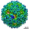
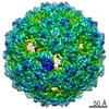
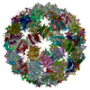
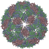
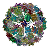
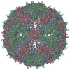
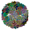
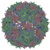
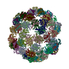
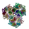
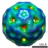
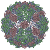
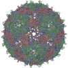
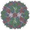
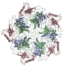
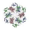
Journal: Proc Natl Acad Sci U S A / Year: 2016 Title: Asymmetric cryo-EM structure of the canonical Allolevivirus Qβ reveals a single maturation protein and the genomic ssRNA in situ. Authors: Karl V Gorzelnik / Zhicheng Cui / Catrina A Reed / Joanita Jakana / Ry Young / Junjie Zhang / Abstract: Single-stranded (ss) RNA viruses infect all domains of life. To date, for most ssRNA virions, only the structures of the capsids and their associated protein components have been resolved to high ...Single-stranded (ss) RNA viruses infect all domains of life. To date, for most ssRNA virions, only the structures of the capsids and their associated protein components have been resolved to high resolution. Qβ, an ssRNA phage specific for the conjugative F-pilus, has a T = 3 icosahedral lattice of coat proteins assembled around its 4,217 nucleotides of genomic RNA (gRNA). In the mature virion, the maturation protein, A, binds to the gRNA and is required for adsorption to the F-pilus. Here, we report the cryo-electron microscopy (cryo-EM) structures of Qβ with and without symmetry applied. The icosahedral structure, at 3.7-Å resolution, resolves loops not previously seen in the published X-ray structure, whereas the asymmetric structure, at 7-Å resolution, reveals A and the gRNA. A contains a bundle of α-helices and replaces one dimer of coat proteins at a twofold axis. The helix bundle binds gRNA, causing denser packing of RNA in its proximity, which asymmetrically expands the surrounding coat protein shell to potentially facilitate RNA release during infection. We observe a fixed pattern of gRNA organization among all viral particles, with the major and minor grooves of RNA helices clearly visible. A single layer of RNA directly contacts every copy of the coat protein, with one-third of the interactions occurring at operator-like RNA hairpins. These RNA-coat interactions stabilize the tertiary structure of gRNA within the virion, which could further provide a roadmap for capsid assembly.
In the structure databanks used in Yorodumi, some data are registered as the other names, "COVID-19 virus" and "2019-nCoV". Here are the details of the virus and the list of structure data.
Jan 31, 2019. EMDB accession codes are about to change! (news from PDBe EMDB page)
EMDB accession codes are about to change! (news from PDBe EMDB page)
The allocation of 4 digits for EMDB accession codes will soon come to an end. Whilst these codes will remain in use, new EMDB accession codes will include an additional digit and will expand incrementally as the available range of codes is exhausted. The current 4-digit format prefixed with “EMD-” (i.e. EMD-XXXX) will advance to a 5-digit format (i.e. EMD-XXXXX), and so on. It is currently estimated that the 4-digit codes will be depleted around Spring 2019, at which point the 5-digit format will come into force.
The EM Navigator/Yorodumi systems omit the EMD- prefix.
Related info.:Q: What is EMD? / ID/Accession-code notation in Yorodumi/EM Navigator
Yorodumi is a browser for structure data from EMDB, PDB, SASBDB, etc.
This page is also the successor to EM Navigator detail page, and also detail information page/front-end page for Omokage search.
The word "yorodu" (or yorozu) is an old Japanese word meaning "ten thousand". "mi" (miru) is to see.
Related info.:EMDB / PDB / SASBDB / Comparison of 3 databanks / Yorodumi Search / Aug 31, 2016. New EM Navigator & Yorodumi / Yorodumi Papers / Jmol/JSmol / Function and homology information / Changes in new EM Navigator and Yorodumi
 Movie
Movie Controller


 Open data
 Basic information
 Components
 Keywords
 Function and homology information
 Enterobacteria phage Qbeta (virus)
 Authors
 United States, 3items
  Citation
 Structure visualization
 Downloads & links
 Other downloads
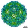
 PDBj
PDBj
 Assembly
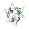

 Sample preparation
 Electron microscopy imaging
 FIELD EMISSION GUN / Accelerating voltage: 300 kV / Illumination mode: FLOOD BEAM
 Processing