 Movie
Movie Controller

[English] 日本語
 Yorodumi
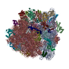
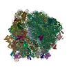
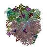
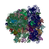
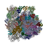
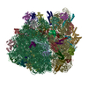
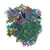
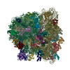
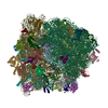
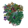
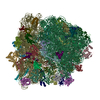
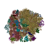
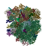
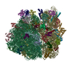
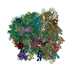
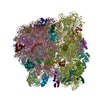
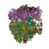
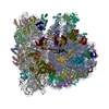
- PDB-5afi: 2.9A Structure of E. coli ribosome-EF-TU complex by cs-corrected ... -
+ Open data

- Basic information
| Entry | Database: PDB / ID: 5afi | |||||||||||||||
|---|---|---|---|---|---|---|---|---|---|---|---|---|---|---|---|---|
| Title | 2.9A Structure of E. coli ribosome-EF-TU complex by cs-corrected cryo-EM | |||||||||||||||
 Components Components |
| |||||||||||||||
 Keywords Keywords | RIBOSOME / TRANSLATION / PROTEIN SYNTHESIS / DECODING / ELONGATION FACTOR TU / TRNA / RNA MODIFICATION / ANTIBIOTIC | |||||||||||||||
| Function / homology |  Function and homology information Function and homology informationguanyl-nucleotide exchange factor complex / protein-synthesizing GTPase / guanosine tetraphosphate binding / stringent response / negative regulation of cytoplasmic translational initiation / transcription antitermination factor activity, RNA binding / ornithine decarboxylase inhibitor activity / translational elongation / misfolded RNA binding / Group I intron splicing ...guanyl-nucleotide exchange factor complex / protein-synthesizing GTPase / guanosine tetraphosphate binding / stringent response / negative regulation of cytoplasmic translational initiation / transcription antitermination factor activity, RNA binding / ornithine decarboxylase inhibitor activity / translational elongation / misfolded RNA binding / Group I intron splicing / RNA folding / translation elongation factor activity / translational termination / transcriptional attenuation / endoribonuclease inhibitor activity / positive regulation of ribosome biogenesis / RNA-binding transcription regulator activity / four-way junction DNA binding / negative regulation of cytoplasmic translation / DnaA-L2 complex / regulation of mRNA stability / translation repressor activity / negative regulation of translational initiation / negative regulation of DNA-templated DNA replication initiation / mRNA regulatory element binding translation repressor activity / positive regulation of RNA splicing / regulation of DNA-templated transcription elongation / transcription elongation factor complex / response to reactive oxygen species / cytosolic ribosome assembly / ribosome assembly / assembly of large subunit precursor of preribosome / transcription antitermination / DNA endonuclease activity / translational initiation / regulation of cell growth / DNA-templated transcription termination / response to radiation / maintenance of translational fidelity / mRNA 5'-UTR binding / regulation of translation / large ribosomal subunit / transferase activity / ribosomal small subunit assembly / ribosome biogenesis / ribosome binding / ribosomal small subunit biogenesis / 5S rRNA binding / ribosomal large subunit assembly / small ribosomal subunit / small ribosomal subunit rRNA binding / large ribosomal subunit rRNA binding / cytosolic small ribosomal subunit / cytosolic large ribosomal subunit / cytoplasmic translation / tRNA binding / negative regulation of translation / rRNA binding / structural constituent of ribosome / ribosome / translation / response to antibiotic / negative regulation of DNA-templated transcription / hydrolase activity / mRNA binding / GTPase activity / GTP binding / magnesium ion binding / DNA binding / RNA binding / zinc ion binding / membrane / plasma membrane / cytoplasm / cytosol Similarity search - Function | |||||||||||||||
| Biological species |  | |||||||||||||||
| Method | ELECTRON MICROSCOPY / single particle reconstruction / cryo EM / Resolution: 2.9 Å | |||||||||||||||
 Authors Authors | Fischer, N. / Neumann, P. / Konevega, A.L. / Bock, L.V. / Ficner, R. / Rodnina, M.V. / Stark, H. | |||||||||||||||
 Citation Citation | Journal: Nature / Year: 2015 Title: Structure of the E. coli ribosome-EF-Tu complex at <3 Å resolution by Cs-corrected cryo-EM. Authors: Niels Fischer / Piotr Neumann / Andrey L Konevega / Lars V Bock / Ralf Ficner / Marina V Rodnina / Holger Stark /  Abstract: Single particle electron cryomicroscopy (cryo-EM) has recently made significant progress in high-resolution structure determination of macromolecular complexes due to improvements in electron ...Single particle electron cryomicroscopy (cryo-EM) has recently made significant progress in high-resolution structure determination of macromolecular complexes due to improvements in electron microscopic instrumentation and computational image analysis. However, cryo-EM structures can be highly non-uniform in local resolution and all structures available to date have been limited to resolutions above 3 Å. Here we present the cryo-EM structure of the 70S ribosome from Escherichia coli in complex with elongation factor Tu, aminoacyl-tRNA and the antibiotic kirromycin at 2.65-2.9 Å resolution using spherical aberration (Cs)-corrected cryo-EM. Overall, the cryo-EM reconstruction at 2.9 Å resolution is comparable to the best-resolved X-ray structure of the E. coli 70S ribosome (2.8 Å), but provides more detailed information (2.65 Å) at the functionally important ribosomal core. The cryo-EM map elucidates for the first time the structure of all 35 rRNA modifications in the bacterial ribosome, explaining their roles in fine-tuning ribosome structure and function and modulating the action of antibiotics. We also obtained atomic models for flexible parts of the ribosome such as ribosomal proteins L9 and L31. The refined cryo-EM-based model presents the currently most complete high-resolution structure of the E. coli ribosome, which demonstrates the power of cryo-EM in structure determination of large and dynamic macromolecular complexes. | |||||||||||||||
| History |
|
- Structure visualization
| Movie |
Movie viewer |
|---|---|
| Structure viewer | Molecule: MolmilJmol/JSmol |
 UCSF Chimera
- Downloads & links
-Download
| PDBx/mmCIF format | 5afi.cif.gz | 6.1 MB | Display | PDBx/mmCIF format |
|---|---|---|---|---|
| PDB format | pdb5afi.ent.gz | Display | PDB format | |
| PDBx/mmJSON format | 5afi.json.gz | Tree view | PDBx/mmJSON format | |
| Others |  Other downloads Other downloads |
-Validation report
| Arichive directory | https://data.pdbj.org/pub/pdb/validation_reports/af/5afiftp://data.pdbj.org/pub/pdb/validation_reports/af/5afi | HTTPS FTP |
|---|
-Related structure data
| Related structure data | 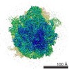 2847MC M: map data used to model this data C: citing same article ( |
|---|---|
| Similar structure data |

-Links
 PDBj
PDBj


- Assembly
| Deposited unit | 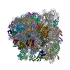
|
|---|---|
| 1 |
|
-Components
-RNA chain , 6 types, 7 molecules avwxyAB
| #1: RNA chain | Mass: 498909.844 Da / Num. of mol.: 1 / Source method: isolated from a natural source / Source: (natural) | ||||||||
|---|---|---|---|---|---|---|---|---|---|
| #22: RNA chain | Mass: 24818.893 Da / Num. of mol.: 2 / Fragment: Residues 49-125 / Source method: isolated from a natural source / Source: (natural) #23: RNA chain | | Mass: 3492.122 Da / Num. of mol.: 1 / Source method: isolated from a natural source / Source: (natural) #24: RNA chain | | Mass: 24797.088 Da / Num. of mol.: 1 / Source method: isolated from a natural source / Source: (natural) #26: RNA chain | | Mass: 941521.375 Da / Num. of mol.: 1 / Source method: isolated from a natural source / Source: (natural) #27: RNA chain | | Mass: 38813.133 Da / Num. of mol.: 1 / Source method: isolated from a natural source / Source: (natural) |
-30S ribosomal protein ... , 20 types, 20 molecules bcdefghijklmnopqrstu
| #2: Protein | Mass: 26652.557 Da / Num. of mol.: 1 / Source method: isolated from a natural source / Source: (natural) |
|---|---|
| #3: Protein | Mass: 26031.316 Da / Num. of mol.: 1 / Source method: isolated from a natural source / Source: (natural) |
| #4: Protein | Mass: 23514.199 Da / Num. of mol.: 1 / Source method: isolated from a natural source / Source: (natural) |
| #5: Protein | Mass: 17629.398 Da / Num. of mol.: 1 / Source method: isolated from a natural source / Source: (natural) |
| #6: Protein | Mass: 15727.512 Da / Num. of mol.: 1 / Source method: isolated from a natural source / Source: (natural) |
| #7: Protein | Mass: 20055.156 Da / Num. of mol.: 1 / Source method: isolated from a natural source / Source: (natural) |
| #8: Protein | Mass: 14146.557 Da / Num. of mol.: 1 / Source method: isolated from a natural source / Source: (natural) |
| #9: Protein | Mass: 14886.270 Da / Num. of mol.: 1 / Source method: isolated from a natural source / Source: (natural) |
| #10: Protein | Mass: 11755.597 Da / Num. of mol.: 1 / Source method: isolated from a natural source / Source: (natural) |
| #11: Protein | Mass: 13870.975 Da / Num. of mol.: 1 / Source method: isolated from a natural source / Source: (natural) |
| #12: Protein | Mass: 13768.157 Da / Num. of mol.: 1 / Source method: isolated from a natural source / Source: (natural) |
| #13: Protein | Mass: 13128.467 Da / Num. of mol.: 1 / Source method: isolated from a natural source / Source: (natural) |
| #14: Protein | Mass: 11677.637 Da / Num. of mol.: 1 / Source method: isolated from a natural source / Source: (natural) |
| #15: Protein | Mass: 10290.816 Da / Num. of mol.: 1 / Source method: isolated from a natural source / Source: (natural) |
| #16: Protein | Mass: 9207.572 Da / Num. of mol.: 1 / Source method: isolated from a natural source / Source: (natural) |
| #17: Protein | Mass: 9724.491 Da / Num. of mol.: 1 / Source method: isolated from a natural source / Source: (natural) |
| #18: Protein | Mass: 9005.472 Da / Num. of mol.: 1 / Source method: isolated from a natural source / Source: (natural) |
| #19: Protein | Mass: 10455.355 Da / Num. of mol.: 1 / Source method: isolated from a natural source / Source: (natural) |
| #20: Protein | Mass: 9708.464 Da / Num. of mol.: 1 / Source method: isolated from a natural source / Source: (natural) |
| #21: Protein | Mass: 8524.039 Da / Num. of mol.: 1 / Source method: isolated from a natural source / Source: (natural) |
-Protein , 1 types, 1 molecules z
| #25: Protein | Mass: 43239.297 Da / Num. of mol.: 1 / Source method: isolated from a natural source / Source: (natural) |
|---|
+50S ribosomal protein ... , 31 types, 31 molecules CDEFGHIJKLMNOPQRSTUVWXYZ0123456
-Non-polymers , 8 types, 363 molecules 


| #59: Chemical | ChemComp-MG /  Mass: 24.305 Da / Num. of mol.: 333 / Source method: obtained synthetically / Formula: Mg / References: UniProt: P0A7M9*PLUS Mass: 24.305 Da / Num. of mol.: 333 / Source method: obtained synthetically / Formula: Mg / References: UniProt: P0A7M9*PLUS#60: Chemical |  Mass: 35.453 Da / Num. of mol.: 2 / Source method: obtained synthetically / Formula: Cl Mass: 35.453 Da / Num. of mol.: 2 / Source method: obtained synthetically / Formula: Cl#61: Chemical | ChemComp-FME / |  Type: L-peptide linking / Mass: 177.221 Da / Num. of mol.: 1 / Source method: isolated from a natural source / Formula: C6H11NO3S / Source: (natural) Type: L-peptide linking / Mass: 177.221 Da / Num. of mol.: 1 / Source method: isolated from a natural source / Formula: C6H11NO3S / Source: (natural) #62: Chemical | ChemComp-KIR / |  Mass: 796.943 Da / Num. of mol.: 1 / Source method: obtained synthetically / Formula: C43H60N2O12 Mass: 796.943 Da / Num. of mol.: 1 / Source method: obtained synthetically / Formula: C43H60N2O12#63: Chemical | ChemComp-GDP / |  Type: RNA linking / Mass: 443.201 Da / Num. of mol.: 1 / Source method: obtained synthetically / Formula: C10H15N5O11P2 / Comment: GDP, energy-carrying molecule*YM Type: RNA linking / Mass: 443.201 Da / Num. of mol.: 1 / Source method: obtained synthetically / Formula: C10H15N5O11P2 / Comment: GDP, energy-carrying molecule*YM#64: Chemical |  Mass: 22.990 Da / Num. of mol.: 2 / Source method: obtained synthetically / Formula: Na Mass: 22.990 Da / Num. of mol.: 2 / Source method: obtained synthetically / Formula: Na#65: Chemical |  Mass: 65.409 Da / Num. of mol.: 2 / Source method: obtained synthetically / Formula: Zn Mass: 65.409 Da / Num. of mol.: 2 / Source method: obtained synthetically / Formula: Zn#66: Water | ChemComp-HOH / | Mass: 18.015 Da / Num. of mol.: 21 / Source method: isolated from a natural source / Formula: H2O |
|---|
-Details
| Has protein modification | Y |
|---|
-Experimental details
-Experiment
| Experiment | Method: ELECTRON MICROSCOPY |
|---|---|
| EM experiment | Aggregation state: PARTICLE / 3D reconstruction method: single particle reconstruction |
- Sample preparation
| Component | Name: E. COLI 70S-EF-TU-GDP- KIRROMYCIN-PHE-TRNAPHE- FMET-TRNAFMET-TRNAFMET COMPLEX Type: RIBOSOME |
|---|---|
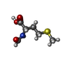
| Buffer solution | Name: 50MM HEPES-KOH, 70MMNH4CL, 30MM KCL, 20MM MGCL2, 1MM DTT, 0.6MM SPERMINE, 0.4MM SPERMIDINE, 0.15MM KIRROMYCIN pH: 7.5 Details: 50MM HEPES-KOH, 70MMNH4CL, 30MM KCL, 20MM MGCL2, 1MM DTT, 0.6MM SPERMINE, 0.4MM SPERMIDINE, 0.15MM KIRROMYCIN |
| Specimen | Embedding applied: NO / Shadowing applied: NO / Staining applied: NO / Vitrification applied: YES |
| Specimen support | Details: CARBON |
| Vitrification | Instrument: FEI VITROBOT MARK IV / Cryogen name: ETHANE / Details: LIQUIDE ETHANE |
- Electron microscopy imaging
| Experimental equipment |  Model: Titan Krios / Image courtesy: FEI Company |
|---|---|
| Microscopy | Model: FEI TITAN KRIOS / Date: Dec 20, 2011 |
| Electron gun | Electron source: OTHER / Accelerating voltage: 300 kV / Illumination mode: SPOT SCAN |
| Electron lens | Mode: BRIGHT FIELD / Nominal magnification: 192000 X / Nominal defocus max: 2500 nm / Nominal defocus min: 700 nm / Cs: 0.01 mm |
| Image recording | Electron dose: 40 e/Å2 / Film or detector model: FEI FALCON I (4k x 4k) |
- Processing
| EM software |
| ||||||||||||||||
|---|---|---|---|---|---|---|---|---|---|---|---|---|---|---|---|---|---|
| CTF correction | Details: LOCAL CTF CORRECTION | ||||||||||||||||
| Symmetry | Point symmetry: C1 (asymmetric) | ||||||||||||||||
| 3D reconstruction | Resolution: 2.9 Å / Num. of particles: 417201 / Actual pixel size: 0.75525 Å Magnification calibration: CROSS- -CORRELATION WITH PUBLISHED ATOMIC COORDINATES Symmetry type: POINT | ||||||||||||||||
| Atomic model building | Space: REAL | ||||||||||||||||
| Refinement | Highest resolution: 2.9 Å |
