 Movie
Movie Controller


+ Open data

- Basic information
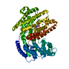
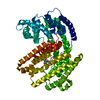
| Entry | Database: PDB / ID: 5eau | ||||||
|---|---|---|---|---|---|---|---|
| Title | 5-EPI-ARISTOLOCHENE SYNTHASE FROM NICOTIANA TABACUM | ||||||
 Components Components | 5-EPI-ARISTOLOCHENE SYNTHASE | ||||||
 Keywords Keywords | ISOPRENOID SYNTHASE / 5-EPI-ARISTOLOCHENE SYNTHASE / NATURAL PRODUCTS BIOSYNTHESIS / ISOPRENOID CYCLASE | ||||||
| Function / homology |  Function and homology information Function and homology information(+)-2-epi-prezizaene synthase / (-)-alpha-cedrene synthase / 5-epiaristolochene synthase / terpene biosynthetic process / 5-epi-aristolochene synthase activity / sesquiterpene biosynthetic process / diterpenoid biosynthetic process / terpene synthase activity / magnesium ion binding / cytoplasm Similarity search - Function | ||||||
| Biological species |  | ||||||
| Method |  X-RAY DIFFRACTION / SYNCHROTRON / DIFFERENCE FOURIER / Resolution: 2.15 Å X-RAY DIFFRACTION / SYNCHROTRON / DIFFERENCE FOURIER / Resolution: 2.15 Å | ||||||
 Authors Authors | Starks, C.M. / Back, K. / Chappell, J. / Noel, J.P. | ||||||
 Citation Citation | Journal: Science / Year: 1997 Title: Structural basis for cyclic terpene biosynthesis by tobacco 5-epi-aristolochene synthase. Authors: Starks, C.M. / Back, K. / Chappell, J. / Noel, J.P. #1: Journal: Arch.Biochem.Biophys. / Year: 1994Title: Expression of a Plant Sesquiterpene Cyclase Gene in Escherichia Coli Authors: Back, K. / Yin, S. / Chappell, J. #2: Journal: Plant Physiol. / Year: 1993Title: Purification and Characterization of an Inducible Sesquiterpene Cyclase from Elicitor-Treated Tobacco Cell Suspension Cultures Authors: Vogeli, U. / Freeman, J.W. / Chappell, J. | ||||||
| History |
|
- Structure visualization
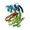
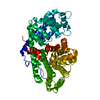
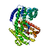
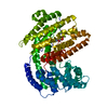
| Structure viewer | Molecule: MolmilJmol/JSmol |
|---|
- Downloads & links
-Download
| PDBx/mmCIF format | 5eau.cif.gz | 120.9 KB | Display | PDBx/mmCIF format |
|---|---|---|---|---|
| PDB format | pdb5eau.ent.gz | 93.9 KB | Display | PDB format |
| PDBx/mmJSON format | 5eau.json.gz | Tree view | PDBx/mmJSON format | |
| Others |  Other downloads Other downloads |
-Validation report
| Arichive directory | https://data.pdbj.org/pub/pdb/validation_reports/ea/5eauftp://data.pdbj.org/pub/pdb/validation_reports/ea/5eau | HTTPS FTP |
|---|
-Related structure data
| Related structure data |  5easSC  5eatC S: Starting model for refinement C: citing same article ( |
|---|---|
| Similar structure data |

-Links
 PDBj
PDBj

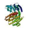
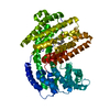
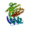
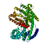
- Assembly
| Deposited unit | 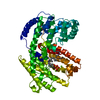
| ||||||||
|---|---|---|---|---|---|---|---|---|---|
| 1 |
| ||||||||
| Unit cell |
|
-Components
| #1: Protein | Mass: 63043.484 Da / Num. of mol.: 1 / Mutation: EXPRESSED WITH 6-HIS TAG Source method: isolated from a genetically manipulated source Source: (gene. exp.)  | ||||
|---|---|---|---|---|---|
| #2: Chemical |   Mass: 24.305 Da / Num. of mol.: 2 / Source method: obtained synthetically / Formula: Mg Mass: 24.305 Da / Num. of mol.: 2 / Source method: obtained synthetically / Formula: Mg#3: Chemical | ChemComp-FFF / | 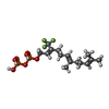  Mass: 436.298 Da / Num. of mol.: 1 / Source method: obtained synthetically / Formula: C15H25F3O7P2 Mass: 436.298 Da / Num. of mol.: 1 / Source method: obtained synthetically / Formula: C15H25F3O7P2#4: Water | ChemComp-HOH / |  Mass: 18.015 Da / Num. of mol.: 131 / Source method: isolated from a natural source / Formula: H2O Mass: 18.015 Da / Num. of mol.: 131 / Source method: isolated from a natural source / Formula: H2O |
-Experimental details
-Experiment
| Experiment | Method: X-RAY DIFFRACTION / Number of used crystals: 1 |
|---|
- Sample preparation
| Crystal | Density Matthews: 3.87 Å3/Da / Density % sol: 68 % | ||||||||||||||||||||||||||||||
|---|---|---|---|---|---|---|---|---|---|---|---|---|---|---|---|---|---|---|---|---|---|---|---|---|---|---|---|---|---|---|---|
| Crystal grow | pH: 6.9 Details: PROTEIN WAS CRYSTALLIZED FROM 15% PEG 8000, 200 MM MG(OAC)2, 100 MM MOPSO PH 6.9, 1MM DTT; SOAKING SOLUTION FOR FREEZING ALSO INCLUDED 20% ETHYLENE GLYCOL. | ||||||||||||||||||||||||||||||
| Crystal | *PLUS | ||||||||||||||||||||||||||||||
| Crystal grow | *PLUS Method: vapor diffusion, hanging drop | ||||||||||||||||||||||||||||||
| Components of the solutions | *PLUS
|
-Data collection
| Diffraction | Mean temperature: 90 K |
|---|---|
| Diffraction source | Source: SYNCHROTRON / Site: SSRL  / Beamline: BL7-1 / Wavelength: 1.08 / Beamline: BL7-1 / Wavelength: 1.08 |
| Detector | Type: MARRESEARCH / Detector: IMAGE PLATE / Date: Mar 1, 1996 |
| Radiation | Monochromatic (M) / Laue (L): M / Scattering type: x-ray |
| Radiation wavelength | Wavelength: 1.08 Å / Relative weight: 1 |
| Reflection | Resolution: 2.16→23.3 Å / Num. obs: 49688 / % possible obs: 91.5 % / Observed criterion σ(I): -2 / Redundancy: 5.4 % / Rsym value: 0.052 / Net I/σ(I): 8 |
| Reflection shell | Resolution: 2.15→2.2 Å / Redundancy: 3.8 % / Mean I/σ(I) obs: 2.7 / Rsym value: 0.444 / % possible all: 70.3 |
| Reflection | *PLUS Num. measured all: 268076 / Rmerge(I) obs: 0.076 |
| Reflection shell | *PLUS % possible obs: 70.3 % / Rmerge(I) obs: 0.469 |
- Processing
| Software |
| ||||||||||||||||||||||||||||||||||||||||||||||||||||||||||||
|---|---|---|---|---|---|---|---|---|---|---|---|---|---|---|---|---|---|---|---|---|---|---|---|---|---|---|---|---|---|---|---|---|---|---|---|---|---|---|---|---|---|---|---|---|---|---|---|---|---|---|---|---|---|---|---|---|---|---|---|---|---|
| Refinement | Method to determine structure: DIFFERENCE FOURIER Starting model: PDB ENTRY 5EAS Resolution: 2.15→22 Å / Rfactor Rfree error: 0.006 / Data cutoff high absF: 1000000 / Data cutoff low absF: 0.001 / Isotropic thermal model: RESTRAINED / Cross valid method: THROUGHOUT Details: THE PROTEIN CONSISTS OF RESIDUES 1 - 548. RESIDUES 1 - 20 ARE ABSENT FROM THE ELECTRON DENSITY. RESIDUES 97 - 102 EXHIBIT WEAK DENSITY, AND MAY BE IN MULTIPLE CONFORMATIONS. RESIDUES 315 - ...Details: THE PROTEIN CONSISTS OF RESIDUES 1 - 548. RESIDUES 1 - 20 ARE ABSENT FROM THE ELECTRON DENSITY. RESIDUES 97 - 102 EXHIBIT WEAK DENSITY, AND MAY BE IN MULTIPLE CONFORMATIONS. RESIDUES 315 - 333 EXHIBIT ANISOTROPIC ELECTRON DENSITY. RESIDUES 524 - 528 ARE ABSENT FROM THE ELECTRON DENSITY. MUCH OF THE FARNESYL CHAIN OF THE BOUND SUBSTRATE ANALOG IS DISORDERED.
| ||||||||||||||||||||||||||||||||||||||||||||||||||||||||||||
| Displacement parameters | Biso mean: 40.7 Å2 | ||||||||||||||||||||||||||||||||||||||||||||||||||||||||||||
| Refinement step | Cycle: LAST / Resolution: 2.15→22 Å
| ||||||||||||||||||||||||||||||||||||||||||||||||||||||||||||
| Refine LS restraints |
| ||||||||||||||||||||||||||||||||||||||||||||||||||||||||||||
| LS refinement shell | Resolution: 2.15→2.25 Å / Rfactor Rfree error: 0.03 / Total num. of bins used: 8
| ||||||||||||||||||||||||||||||||||||||||||||||||||||||||||||
| Xplor file |
| ||||||||||||||||||||||||||||||||||||||||||||||||||||||||||||
| Software | *PLUS Name: X-PLOR / Version: 3.843 / Classification: refinement | ||||||||||||||||||||||||||||||||||||||||||||||||||||||||||||
| Refine LS restraints | *PLUS
|
