ムービー
ムービー コントローラー


+ データを開く

- 基本情報
基本情報
| 登録情報 | データベース: EMDB / ID: EMD-5529 | |||||||||
|---|---|---|---|---|---|---|---|---|---|---|
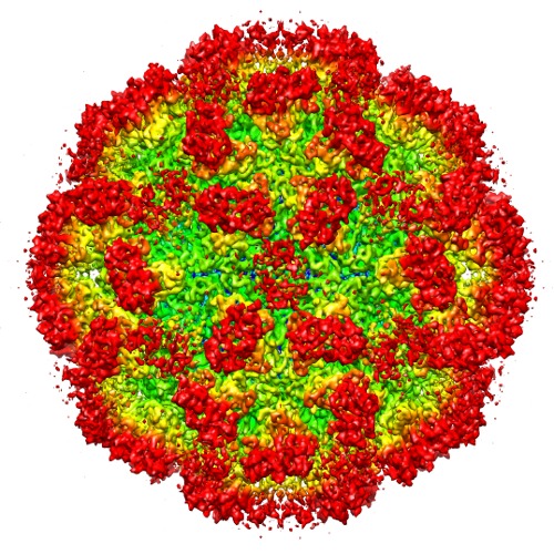
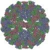
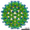
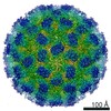
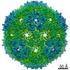
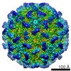
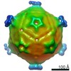
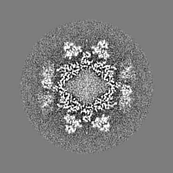
| タイトル | 6.3 A Cryo-EM Structure of a Novel Calicivirus, Tulane Virus | |||||||||
 マップデータ マップデータ | Reconstruction of TV virion | |||||||||
 試料 試料 |
| |||||||||
 キーワード キーワード | Tulane virus / calicivirus / conformational flexibility / single particle cryo-EM / 3-D reconstruction | |||||||||
| 生物種 |  Tulane virus (ウイルス) Tulane virus (ウイルス) | |||||||||
| 手法 | 単粒子再構成法 / クライオ電子顕微鏡法 / ネガティブ染色法 / 解像度: 6.3 Å | |||||||||
 データ登録者 データ登録者 | Yu G / Zhang D / Guo F / Tan M / Jiang X / Jiang W | |||||||||
 引用 引用 | ジャーナル: PLoS One / 年: 2013 タイトル: Cryo-EM structure of a novel calicivirus, Tulane virus. 著者: Guimei Yu / Dongsheng Zhang / Fei Guo / Ming Tan / Xi Jiang / Wen Jiang /  要旨: Tulane virus (TV) is a newly isolated cultivatable calicivirus that infects juvenile rhesus macaques. Here we report a 6.3 Å resolution cryo-electron microscopy structure of the TV virion. The TV ...Tulane virus (TV) is a newly isolated cultivatable calicivirus that infects juvenile rhesus macaques. Here we report a 6.3 Å resolution cryo-electron microscopy structure of the TV virion. The TV virion is about 400 Å in diameter and consists of a T = 3 icosahedral protein capsid enclosing the RNA genome. 180 copies of the major capsid protein VP1 (∼57 KDa) are organized into two types of dimers A/B and C/C and form a thin, smooth shell studded with 90 dimeric protrusions. The overall capsid organization and the capsid protein fold of TV closely resemble that of other caliciviruses, especially of human Norwalk virus, the prototype human norovirus. These close structural similarities support TV as an attractive surrogate for the non-cultivatable human noroviruses. The most distinctive feature of TV is that its C/C dimers are in a highly flexible conformation with significantly reduced interactions between the shell (S) domain and the protruding (P) domain of VP1. A comparative structural analysis indicated that the P domains of TV C/C dimers were much more flexible than those of other caliciviruses. These observations, combined with previous studies on other caliciviruses, led us to hypothesize that the enhanced flexibility of C/C dimer P domains are likely required for efficient calicivirus-host cell interactions and the consequent uncoating and genome release. Residues in the S-P1 hinge between the S and P domain may play a critical role in the flexibility of P domains of C/C dimers. | |||||||||
| 履歴 |
|
- 構造の表示
構造の表示
| ムービー |
 ムービービューア ムービービューア |
|---|---|
| 構造ビューア | EMマップ: SurfViewMolmilJmol/JSmol |
| 添付画像 |
- ダウンロードとリンク
-EMDBアーカイブ
| マップデータ | emd_5529.map.gz | 55.9 MB | EMDBマップデータ形式 | |
|---|---|---|---|---|
| ヘッダ (付随情報) | emd-5529-v30.xmlemd-5529.xml | 12.4 KB 12.4 KB | 表示 表示 | EMDBヘッダ |
| 画像 | emd_5529.tif | 732.8 KB | ||
| アーカイブディレクトリ |  http://ftp.pdbj.org/pub/emdb/structures/EMD-5529ftp://ftp.pdbj.org/pub/emdb/structures/EMD-5529 http://ftp.pdbj.org/pub/emdb/structures/EMD-5529ftp://ftp.pdbj.org/pub/emdb/structures/EMD-5529 | HTTPS FTP |
-検証レポート
| 文書・要旨 | emd_5529_validation.pdf.gz | 79.3 KB | 表示 | EMDB検証レポート |
|---|---|---|---|---|
| 文書・詳細版 | emd_5529_full_validation.pdf.gz | 78.4 KB | 表示 | |
| XML形式データ | emd_5529_validation.xml.gz | 493 B | 表示 | |
| アーカイブディレクトリ | https://ftp.pdbj.org/pub/emdb/validation_reports/EMD-5529ftp://ftp.pdbj.org/pub/emdb/validation_reports/EMD-5529 | HTTPS FTP |
-関連構造データ
-リンク
| EMDBのページ | EMDB (EBI/PDBe) / EMDataResource |
|---|
-マップ
| ファイル | ダウンロード / ファイル: emd_5529.map.gz / 形式: CCP4 / 大きさ: 162.5 MB / タイプ: IMAGE STORED AS FLOATING POINT NUMBER (4 BYTES) | ||||||||||||||||||||||||||||||||||||||||||||||||||||||||||||||||||||
|---|---|---|---|---|---|---|---|---|---|---|---|---|---|---|---|---|---|---|---|---|---|---|---|---|---|---|---|---|---|---|---|---|---|---|---|---|---|---|---|---|---|---|---|---|---|---|---|---|---|---|---|---|---|---|---|---|---|---|---|---|---|---|---|---|---|---|---|---|---|
| 注釈 | Reconstruction of TV virion | ||||||||||||||||||||||||||||||||||||||||||||||||||||||||||||||||||||
| 投影像・断面図 | 画像のコントロール
画像は Spider により作成 | ||||||||||||||||||||||||||||||||||||||||||||||||||||||||||||||||||||
| ボクセルのサイズ | X=Y=Z: 1.74 Å | ||||||||||||||||||||||||||||||||||||||||||||||||||||||||||||||||||||
| 密度 |
| ||||||||||||||||||||||||||||||||||||||||||||||||||||||||||||||||||||
| 対称性 | 空間群: 1 | ||||||||||||||||||||||||||||||||||||||||||||||||||||||||||||||||||||
| 詳細 | EMDB XML:
CCP4マップ ヘッダ情報:
| ||||||||||||||||||||||||||||||||||||||||||||||||||||||||||||||||||||
 Z (Sec.)
 Y (Row.)
 X (Col.)


-添付データ
- 試料の構成要素
-全体 : Tulane virus
| 全体 | 名称: Tulane virus (ウイルス) |
|---|---|
| 要素 |
|
-超分子 #1000: Tulane virus
| 超分子 | 名称: Tulane virus / タイプ: sample / ID: 1000 集合状態: One Tulane virus has 90 dimers forming its icosahedral capsid (T=3). Number unique components: 1 |
|---|---|
| 分子量 | 理論値: 10.4 MDa |
-超分子 #1: Tulane virus
| 超分子 | 名称: Tulane virus / タイプ: virus / ID: 1 / NCBI-ID: 512169 / 生物種: Tulane virus / データベース: NCBI / ウイルスタイプ: VIRION / ウイルス・単離状態: SPECIES / ウイルス・エンベロープ: No / ウイルス・中空状態: No |
|---|---|
| 宿主 | 生物種:  |
| 分子量 | 理論値: 10.4 MDa |
| ウイルス殻 | Shell ID: 1 / 名称: VP1 / 直径: 400 Å / T番号(三角分割数): 3 |
-実験情報
-構造解析
| 手法 | ネガティブ染色法, クライオ電子顕微鏡法 |
|---|---|
 解析 解析 | 単粒子再構成法 |
| 試料の集合状態 | particle |
-試料調製
| 緩衝液 | pH: 7.4 詳細: 137mM NaCl, 2.7mM KCl, 10mM Na2HPO4, 2mM KH2PO4, pH 7.4 |
|---|---|
| 染色 | タイプ: NEGATIVE 詳細: Grids with sample floated on 2% uranyl acetate for 30 seconds. |
| グリッド | 詳細: 400 mesh copper grid with one lacy carbon layer and one layer of ultra thin carbon on top. |
| 凍結 | 凍結剤: ETHANE / チャンバー内温度: 85 K / 装置: HOMEMADE PLUNGER |
- 電子顕微鏡法
| 顕微鏡 | FEI TITAN KRIOS |
|---|---|
| 温度 | 最低: 80 K / 最高: 85 K / 平均: 80 K |
| アライメント法 | Legacy - 非点収差: Objective lens astigmatism was corrected at 250,000 magnification using quadrupole stigmator. Legacy - Electron beam tilt params: 0 |
| 日付 | 2011年7月29日 |
| 撮影 | カテゴリ: FILM / フィルム・検出器のモデル: KODAK SO-163 FILM デジタル化 - スキャナー: NIKON SUPER COOLSCAN 9000 デジタル化 - サンプリング間隔: 6.35 µm / 実像数: 190 / 平均電子線量: 25 e/Å2 / ビット/ピクセル: 16 |
| 電子線 | 加速電圧: 300 kV / 電子線源:  FIELD EMISSION GUN FIELD EMISSION GUN |
| 電子光学系 | 倍率(補正後): 36475 / 照射モード: FLOOD BEAM / 撮影モード: BRIGHT FIELD / Cs: 2.7 mm / 最大 デフォーカス(公称値): 4.891 µm / 最小 デフォーカス(公称値): 1.347 µm / 倍率(公称値): 37000 |
| 試料ステージ | 試料ホルダー: Liquid nitrogen cooled 試料ホルダーモデル: FEI TITAN KRIOS AUTOGRID HOLDER |
| 実験機器 |  モデル: Titan Krios / 画像提供: FEI Company |
-画像解析
| 詳細 | 4702 Tulane virus particles were selected using combined automated selection with ethan program and manual screening with boxer program in EMAN. The microscope contrast transfer function parameters for each micrograph were first determined using an automated fitting method and then manually verified/corrected using EMAN ctfit graphic program. The entire TV dataset was divided into two halves and processed independently for all the subsequent steps including construction of initial model, 2-D alignment and 3-D reconstruction. De novo initial models were constructed using the random model method in which random particle orientations were assigned and subsequently refined iteratively until convergence. The iterative refinement process including particle alignment and 3-D icosahedral reconstruction was performed using an in-house developed program jspr.py utilizing the EMAN/EMAN2 programs and library functions. The resolution was determined based on the 0.143 cutoff criterion for two truly independent reconstructions. |
|---|---|
| CTF補正 | 詳細: each particle |
| 最終 再構成 | アルゴリズム: OTHER / 解像度のタイプ: BY AUTHOR / 解像度: 6.3 Å / 解像度の算出法: OTHER / ソフトウェア - 名称: jspr.py, EMAN, EMAN2 / 使用した粒子像数: 4338 |
-原子モデル構築 1
| 初期モデル | PDB ID: Chain - #0 - Chain ID: A / Chain - #1 - Chain ID: B / Chain - #2 - Chain ID: C |
|---|---|
| ソフトウェア | 名称: Chimera |
| 詳細 | Protocol: rigid body. The three chains from 1IHM were first fitted into TV density as a whole rigid body and then divided into dimers, specific chains, domains, and subdomains and fitted. |
| 精密化 | 空間: REAL / プロトコル: RIGID BODY FIT / 当てはまり具合の基準: Correlation |