 Movie
Movie Controller


+ Open data

- Basic information
| Entry | Database: EMDB / ID: EMD-5242 | |||||||||
|---|---|---|---|---|---|---|---|---|---|---|
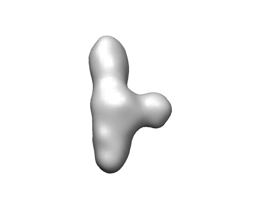
| Title | B. subtilis RNase P RNA Specificity domain folding intermediate | |||||||||
 Map data Map data | This is a map of the folding intermediate of B. subtilis RNase P Specificity domain | |||||||||
 Sample Sample |
| |||||||||
 Keywords Keywords | RNA folding intermediate RNase P Specificity domain | |||||||||
| Biological species |  | |||||||||
| Method | single particle reconstruction / cryo EM / Resolution: 15.2 Å | |||||||||
 Authors Authors | Baird NJ / Ludtke SJ / Khant H / Chiu W / Pan T / Sosnick TR | |||||||||
 Citation Citation | Journal: J Am Chem Soc / Year: 2010 Title: Discrete structure of an RNA folding intermediate revealed by cryo-electron microscopy. Authors: Nathan J Baird / Steven J Ludtke / Htet Khant / Wah Chiu / Tao Pan / Tobin R Sosnick /  Abstract: RNA folding occurs via a series of transitions between metastable intermediate states. It is unknown whether folding intermediates are discrete structures folding along defined pathways or ...RNA folding occurs via a series of transitions between metastable intermediate states. It is unknown whether folding intermediates are discrete structures folding along defined pathways or heterogeneous ensembles folding along broad landscapes. We use cryo-electron microscopy and single-particle image reconstruction to determine the structure of the major folding intermediate of the specificity domain of a ribonuclease P ribozyme. Our results support the existence of a discrete conformation for this folding intermediate. | |||||||||
| History |
|
- Structure visualization
| Movie |
 Movie viewer Movie viewer |
|---|---|
| Structure viewer | EM map: SurfViewMolmilJmol/JSmol |
| Supplemental images |
- Downloads & links
-EMDB archive
| Map data | emd_5242.map.gz | 14.1 MB | EMDB map data format | |
|---|---|---|---|---|
| Header (meta data) | emd-5242-v30.xmlemd-5242.xml | 10.1 KB 10.1 KB | Display Display | EMDB header |
| Images | emd_5242_1.tif | 34.7 KB | ||
| Archive directory |  http://ftp.pdbj.org/pub/emdb/structures/EMD-5242ftp://ftp.pdbj.org/pub/emdb/structures/EMD-5242 http://ftp.pdbj.org/pub/emdb/structures/EMD-5242ftp://ftp.pdbj.org/pub/emdb/structures/EMD-5242 | HTTPS FTP |
-Validation report
| Summary document | emd_5242_validation.pdf.gz | 77.9 KB | Display | EMDB validaton report |
|---|---|---|---|---|
| Full document | emd_5242_full_validation.pdf.gz | 77 KB | Display | |
| Data in XML | emd_5242_validation.xml.gz | 492 B | Display | |
| Arichive directory | https://ftp.pdbj.org/pub/emdb/validation_reports/EMD-5242ftp://ftp.pdbj.org/pub/emdb/validation_reports/EMD-5242 | HTTPS FTP |
-Related structure data


-Links
| EMDB pages | EMDB (EBI/PDBe) / EMDataResource |
|---|
-Map
| File | Download / File: emd_5242.map.gz / Format: CCP4 / Size: 15.3 MB / Type: IMAGE STORED AS FLOATING POINT NUMBER (4 BYTES) | ||||||||||||||||||||||||||||||||||||||||||||||||||||||||||||||||||||
|---|---|---|---|---|---|---|---|---|---|---|---|---|---|---|---|---|---|---|---|---|---|---|---|---|---|---|---|---|---|---|---|---|---|---|---|---|---|---|---|---|---|---|---|---|---|---|---|---|---|---|---|---|---|---|---|---|---|---|---|---|---|---|---|---|---|---|---|---|---|
| Annotation | This is a map of the folding intermediate of B. subtilis RNase P Specificity domain | ||||||||||||||||||||||||||||||||||||||||||||||||||||||||||||||||||||
| Voxel size | X=Y=Z: 1.81 Å | ||||||||||||||||||||||||||||||||||||||||||||||||||||||||||||||||||||
| Density |
| ||||||||||||||||||||||||||||||||||||||||||||||||||||||||||||||||||||
| Symmetry | Space group: 1 | ||||||||||||||||||||||||||||||||||||||||||||||||||||||||||||||||||||
| Details | EMDB XML:
CCP4 map header:
| ||||||||||||||||||||||||||||||||||||||||||||||||||||||||||||||||||||
-Supplemental data
- Sample components
-Entire : B. subtilis RNase P RNA Specificity domain folding intermediate
| Entire | Name: B. subtilis RNase P RNA Specificity domain folding intermediate |
|---|---|
| Components |
|
-Supramolecule #1000: B. subtilis RNase P RNA Specificity domain folding intermediate
| Supramolecule | Name: B. subtilis RNase P RNA Specificity domain folding intermediate type: sample / ID: 1000 / Details: none / Oligomeric state: Monomer of Specificity domain / Number unique components: 1 |
|---|---|
| Molecular weight | Theoretical: 50 KDa / Method: Calculation from nucleotide sequence, 154mer RNA |
-Macromolecule #1: RNA
| Macromolecule | Name: RNA / type: rna / ID: 1 / Name.synonym: RNase P RNA Specificity domain / Classification: OTHER / Structure: OTHER / Synthetic?: No |
|---|---|
| Source (natural) | Organism: |
| Molecular weight | Theoretical: 50 KDa |
| Sequence | String: GCGAGCCUAG CGAAGUCAUA AGCUAGGGCA GUCUUUAGAG GCUGACGGCA GGAAAAAAGC CUACGUCUUC GGAUAUGGCU GAGUAUCCUU GAAAGUGCCA CAGUGACGAA GUCUCACUAG AAAUGGUGAG AGUGGAACGC GGUAAACCCC UCGC |
-Experimental details
-Structure determination
| Method | cryo EM |
|---|---|
 Processing Processing | single particle reconstruction |
| Aggregation state | particle |
-Sample preparation
| Concentration | 1 mg/mL |
|---|---|
| Buffer | pH: 8 / Details: 1 mM MgCl2, 20 mM TrisHCl pH 8 |
| Grid | Details: 400 mesh carbon grid |
| Vitrification | Cryogen name: ETHANE / Chamber humidity: 100 % / Chamber temperature: 77 K / Instrument: FEI VITROBOT MARK III / Details: Vitrification instrument: FEI Vitrobot mark III / Method: 2 blots 1 second each before plunging |
- Electron microscopy
| Microscope | JEOL 2010F |
|---|---|
| Temperature | Min: 93 K / Max: 95 K / Average: 94.1 K |
| Alignment procedure | Legacy - Astigmatism: object astigmatism correction made at 400,000 times magnification |
| Date | Mar 10, 2006 |
| Image recording | Category: CCD / Film or detector model: GATAN ULTRASCAN 4000 (4k x 4k) / Number real images: 100 / Average electron dose: 16 e/Å2 |
| Electron beam | Acceleration voltage: 200 kV / Electron source:  FIELD EMISSION GUN FIELD EMISSION GUN |
| Electron optics | Illumination mode: FLOOD BEAM / Imaging mode: BRIGHT FIELD / Cs: 2 mm / Nominal defocus max: 3.5 µm / Nominal defocus min: 1.0 µm / Nominal magnification: 60000 |
| Sample stage | Specimen holder: single tilt cryo-holder / Specimen holder model: GATAN LIQUID NITROGEN |
-Image processing
| Details | The particles were selected using an automatic selection program and then inspected manually |
|---|---|
| Final reconstruction | Algorithm: OTHER / Resolution.type: BY AUTHOR / Resolution: 15.2 Å / Resolution method: FSC 0.5 CUT-OFF / Software - Name: EMAN Details: FSC gives a resolution of 15.2 A, but the model was low-pass filtered to 26 A, corresponding to the first zero-crossing of the data. CTF correction was not performed. Number images used: 11600 |
| Final two d classification | Number classes: 60 |
