 Movie
Movie Controller

[English] 日本語
 Yorodumi
- PDB-4xs9: Determining the Molecular Basis for Starter Unit Selection During... -
+ Open data

- Basic information
| Entry | Database: PDB / ID: 4xs9 | ||||||
|---|---|---|---|---|---|---|---|
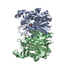
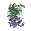
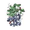
| Title | Determining the Molecular Basis for Starter Unit Selection During Daunorubicin Biosynthesis | ||||||
 Components Components | Daunorubicin-doxorubicin polyketide synthase | ||||||
 Keywords Keywords | TRANSFERASE / polyketide / daunorubicin / natural product / starter unit | ||||||
| Function / homology |  Function and homology information Function and homology informationsecondary metabolite biosynthetic process / acyltransferase activity, transferring groups other than amino-acyl groups Similarity search - Function | ||||||
| Biological species |  Streptomyces peucetius (bacteria) Streptomyces peucetius (bacteria) | ||||||
| Method |  X-RAY DIFFRACTION / SYNCHROTRON / MOLECULAR REPLACEMENT / Resolution: 2.002 Å X-RAY DIFFRACTION / SYNCHROTRON / MOLECULAR REPLACEMENT / Resolution: 2.002 Å | ||||||
 Authors Authors | Jackson, D.R. / Valentic, T.R. / Tsai, S.C. / Patel, A. / Mohammed, L. / Vasilakis, K. / Wattana-amorn, P. / Long, P.F. / Crump, M.P. / Crosby, J. | ||||||
| Funding support |  United States, 1items United States, 1items
| ||||||
 Citation Citation | Journal: To Be Published Title: Determining the Molecular Basis for Starter Unit Selection During Daunorubicin Biosynthesis Authors: Jackson, D.R. / Valentic, T.R. / Tsai, S.C. / Patel, A. / Mohammed, L. / Vasilakis, K. / Wattana-amorn, P. / Long, P.F. / Crump, M.P. / Crosby, J. | ||||||
| History |
|
- Structure visualization
| Structure viewer | Molecule: MolmilJmol/JSmol |
|---|
- Downloads & links
-Download
| PDBx/mmCIF format | 4xs9.cif.gz | 150.6 KB | Display | PDBx/mmCIF format |
|---|---|---|---|---|
| PDB format | pdb4xs9.ent.gz | 116.2 KB | Display | PDB format |
| PDBx/mmJSON format | 4xs9.json.gz | Tree view | PDBx/mmJSON format | |
| Others |  Other downloads Other downloads |
-Validation report
| Arichive directory | https://data.pdbj.org/pub/pdb/validation_reports/xs/4xs9ftp://data.pdbj.org/pub/pdb/validation_reports/xs/4xs9 | HTTPS FTP |
|---|
-Related structure data
| Related structure data |  4xs7C  4xsaC 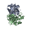 4xsbC  4xqp S: Starting model for refinement C: citing same article ( |
|---|---|
| Similar structure data |


-Links
 PDBj
PDBj

- Assembly
| Deposited unit | 
| ||||||||
|---|---|---|---|---|---|---|---|---|---|
| 1 |
| ||||||||
| Unit cell |
| ||||||||
| Components on special symmetry positions |
|
-Components
| #1: Protein | Mass: 39430.473 Da / Num. of mol.: 2 Source method: isolated from a genetically manipulated source Details: The protein was co-crystallized with a propionyl phosphopantetheine analogue Source: (gene. exp.) Streptomyces peucetius (bacteria) / Gene: dpsC / Plasmid: pET28a / Production host: References: UniProt: Q54816, Transferases; Acyltransferases; Transferring groups other than aminoacyl groups #2: Chemical |   Mass: 397.361 Da / Num. of mol.: 2 / Source method: obtained synthetically / Formula: C14H28N3O8P Mass: 397.361 Da / Num. of mol.: 2 / Source method: obtained synthetically / Formula: C14H28N3O8P#3: Water | ChemComp-HOH / |  Mass: 18.015 Da / Num. of mol.: 435 / Source method: isolated from a natural source / Formula: H2O Mass: 18.015 Da / Num. of mol.: 435 / Source method: isolated from a natural source / Formula: H2O |
|---|
-Experimental details
-Experiment
| Experiment | Method: X-RAY DIFFRACTION |
|---|
- Sample preparation
| Crystal | Density Matthews: 2.24 Å3/Da / Density % sol: 45 % / Description: hexagonal crystals shaped like Monopoly houses |
|---|---|
| Crystal grow | Temperature: 293 K / Method: vapor diffusion, sitting drop / Details: 0.18 M sodium citrate, 26% PEG3350 |
-Data collection
| Diffraction | Mean temperature: 100 K |
|---|---|
| Diffraction source | Source: SYNCHROTRON / Site: SSRL / Beamline: BL12-2 / Wavelength: 0.9795 Å |
| Detector | Type: DECTRIS PILATUS 6M / Detector: PIXEL / Date: Mar 2, 2013 |
| Radiation | Monochromator: double crystal Si(111) / Protocol: SINGLE WAVELENGTH / Monochromatic (M) / Laue (L): M / Scattering type: x-ray |
| Radiation wavelength | Wavelength: 0.9795 Å / Relative weight: 1 |
| Reflection | Resolution: 2→38.88 Å / Num. obs: 49899 / % possible obs: 100 % / Redundancy: 12.8 % / Rmerge(I) obs: 0.137 / Net I/σ(I): 18.2 |
| Reflection shell | Resolution: 2→2.05 Å / Redundancy: 6.4 % / Rmerge(I) obs: 0.903 / Mean I/σ(I) obs: 1.75 / % possible all: 99.9 |
- Processing
| Software |
| |||||||||||||||||||||||||||||||||||||||||||||||||||||||||||||||||||||||||||||||||||||||||||||||||||||||||
|---|---|---|---|---|---|---|---|---|---|---|---|---|---|---|---|---|---|---|---|---|---|---|---|---|---|---|---|---|---|---|---|---|---|---|---|---|---|---|---|---|---|---|---|---|---|---|---|---|---|---|---|---|---|---|---|---|---|---|---|---|---|---|---|---|---|---|---|---|---|---|---|---|---|---|---|---|---|---|---|---|---|---|---|---|---|---|---|---|---|---|---|---|---|---|---|---|---|---|---|---|---|---|---|---|---|---|
| Refinement | Method to determine structure: MOLECULAR REPLACEMENT Starting model: PDB entry 4XQP 4xqp Resolution: 2.002→38.88 Å / SU ML: 0.18 / Cross valid method: FREE R-VALUE / σ(F): 0 / Phase error: 18.46 / Stereochemistry target values: ML
| |||||||||||||||||||||||||||||||||||||||||||||||||||||||||||||||||||||||||||||||||||||||||||||||||||||||||
| Solvent computation | Shrinkage radii: 0.9 Å / VDW probe radii: 1.11 Å / Solvent model: FLAT BULK SOLVENT MODEL | |||||||||||||||||||||||||||||||||||||||||||||||||||||||||||||||||||||||||||||||||||||||||||||||||||||||||
| Refinement step | Cycle: LAST / Resolution: 2.002→38.88 Å
| |||||||||||||||||||||||||||||||||||||||||||||||||||||||||||||||||||||||||||||||||||||||||||||||||||||||||
| Refine LS restraints |
| |||||||||||||||||||||||||||||||||||||||||||||||||||||||||||||||||||||||||||||||||||||||||||||||||||||||||
| LS refinement shell |
|
