 Movie
Movie Controller

[English] 日本語
 Yorodumi
- PDB-4rmd: Crystal structure of human Retinoid X receptor alpha ligand bindi... -
+ Open data

- Basic information
| Entry | Database: PDB / ID: 4rmd | ||||||
|---|---|---|---|---|---|---|---|
| Title | Crystal structure of human Retinoid X receptor alpha ligand binding domain complex with 9cUAB110 and coactivator peptide GRIP-1 | ||||||
 Components Components |
| ||||||
 Keywords Keywords | TRANSCRIPTION / Cancer prevention / ligand binding domain | ||||||
| Function / homology |  Function and homology information Function and homology informationretinoic acid-responsive element binding / NR1H2 & NR1H3 regulate gene expression linked to triglyceride lipolysis in adipose / NR1H2 & NR1H3 regulate gene expression linked to gluconeogenesis / positive regulation of thyroid hormone receptor signaling pathway / NR1H2 & NR1H3 regulate gene expression to limit cholesterol uptake / NR1H2 & NR1H3 regulate gene expression linked to lipogenesis / Carnitine shuttle / retinoic acid binding / positive regulation of vitamin D receptor signaling pathway / TGFBR3 expression ...retinoic acid-responsive element binding / NR1H2 & NR1H3 regulate gene expression linked to triglyceride lipolysis in adipose / NR1H2 & NR1H3 regulate gene expression linked to gluconeogenesis / positive regulation of thyroid hormone receptor signaling pathway / NR1H2 & NR1H3 regulate gene expression to limit cholesterol uptake / NR1H2 & NR1H3 regulate gene expression linked to lipogenesis / Carnitine shuttle / retinoic acid binding / positive regulation of vitamin D receptor signaling pathway / TGFBR3 expression / nuclear vitamin D receptor binding / Signaling by Retinoic Acid / RNA polymerase II intronic transcription regulatory region sequence-specific DNA binding / NR1H2 & NR1H3 regulate gene expression to control bile acid homeostasis / DNA binding domain binding / positive regulation of lipid metabolic process / nuclear steroid receptor activity / LBD domain binding / locomotor rhythm / positive regulation of lipoprotein transport / aryl hydrocarbon receptor binding / cellular response to Thyroglobulin triiodothyronine / regulation of glucose metabolic process / Synthesis of bile acids and bile salts / regulation of lipid metabolic process / monocyte differentiation / Synthesis of bile acids and bile salts via 27-hydroxycholesterol / positive regulation of cholesterol efflux / Endogenous sterols / Synthesis of bile acids and bile salts via 7alpha-hydroxycholesterol / cellular response to low-density lipoprotein particle stimulus / response to retinoic acid / positive regulation of bone mineralization / retinoic acid receptor signaling pathway / Transcriptional regulation of brown and beige adipocyte differentiation by EBF2 / Recycling of bile acids and salts / transcription regulator inhibitor activity / NR1H3 & NR1H2 regulate gene expression linked to cholesterol transport and efflux / cellular response to hormone stimulus / peroxisome proliferator activated receptor signaling pathway / hormone-mediated signaling pathway / Regulation of lipid metabolism by PPARalpha / cell maturation / positive regulation of adipose tissue development / response to progesterone / peptide binding / BMAL1:CLOCK,NPAS2 activates circadian expression / regulation of cellular response to insulin stimulus / RORA,B,C and NR1D1 (REV-ERBA) regulate gene expression / Activation of gene expression by SREBF (SREBP) / SUMOylation of transcription cofactors / Expression of BMAL (ARNTL), CLOCK, and NPAS2 / nuclear receptor binding / transcription coregulator binding / SUMOylation of intracellular receptors / RNA polymerase II transcription regulatory region sequence-specific DNA binding / circadian regulation of gene expression / negative regulation of smoothened signaling pathway / Heme signaling / PPARA activates gene expression / Transcriptional activation of mitochondrial biogenesis / Cytoprotection by HMOX1 / Activated PKN1 stimulates transcription of AR (androgen receptor) regulated genes KLK2 and KLK3 / Nuclear Receptor transcription pathway / Transcriptional regulation of white adipocyte differentiation / mRNA transcription by RNA polymerase II / nuclear receptor activity / RNA polymerase II transcription regulator complex / Activation of anterior HOX genes in hindbrain development during early embryogenesis / Transcriptional regulation of granulopoiesis / sequence-specific double-stranded DNA binding / nervous system development / HATs acetylate histones / MLL4 and MLL3 complexes regulate expression of PPARG target genes in adipogenesis and hepatic steatosis / double-stranded DNA binding / transcription regulator complex / Estrogen-dependent gene expression / sequence-specific DNA binding / DNA-binding transcription factor activity, RNA polymerase II-specific / cell differentiation / transcription coactivator activity / signaling receptor complex / protein dimerization activity / transcription cis-regulatory region binding / nuclear body / RNA polymerase II cis-regulatory region sequence-specific DNA binding / DNA-binding transcription factor activity / protein domain specific binding / chromatin binding / regulation of DNA-templated transcription / positive regulation of DNA-templated transcription / chromatin / enzyme binding / negative regulation of transcription by RNA polymerase II / positive regulation of transcription by RNA polymerase II / protein-containing complex / mitochondrion / zinc ion binding / nucleoplasm / identical protein binding Similarity search - Function | ||||||
| Biological species |  Homo sapiens (human) Homo sapiens (human) | ||||||
| Method |  X-RAY DIFFRACTION / SYNCHROTRON / MOLECULAR REPLACEMENT / Resolution: 1.9 Å X-RAY DIFFRACTION / SYNCHROTRON / MOLECULAR REPLACEMENT / Resolution: 1.9 Å | ||||||
 Authors Authors | Xia, G. / Muccio, D.D. | ||||||
 Citation Citation | Journal: J.Med.Chem. / Year: 2015 Title: Conformationally Defined Rexinoids and Their Efficacy in the Prevention of Mammary Cancers. Authors: Atigadda, V.R. / Xia, G. / Deshpande, A. / Wu, L. / Kedishvili, N. / Smith, C.D. / Krontiras, H. / Bland, K.I. / Grubbs, C.J. / Brouillette, W.J. / Muccio, D.D. | ||||||
| History |
|
- Structure visualization
| Structure viewer | Molecule: MolmilJmol/JSmol |
|---|
- Downloads & links
-Download
| PDBx/mmCIF format | 4rmd.cif.gz | 62.2 KB | Display | PDBx/mmCIF format |
|---|---|---|---|---|
| PDB format | pdb4rmd.ent.gz | 44.4 KB | Display | PDB format |
| PDBx/mmJSON format | 4rmd.json.gz | Tree view | PDBx/mmJSON format | |
| Others |  Other downloads Other downloads |
-Validation report
| Arichive directory | https://data.pdbj.org/pub/pdb/validation_reports/rm/4rmdftp://data.pdbj.org/pub/pdb/validation_reports/rm/4rmd | HTTPS FTP |
|---|
-Related structure data
| Related structure data |  4rfwC  4rmcC  4rmeC  3oapS S: Starting model for refinement C: citing same article ( |
|---|---|
| Similar structure data |

-Links
 PDBj
PDBj


- Assembly
| Deposited unit | 
| ||||||||
|---|---|---|---|---|---|---|---|---|---|
| 1 | 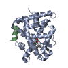
| ||||||||
| Unit cell |
|
-Components
| #1: Protein | Mass: 26395.598 Da / Num. of mol.: 1 / Fragment: ligand binding domain 228-458 Source method: isolated from a genetically manipulated source Source: (gene. exp.) Homo sapiens (human) / Gene: NR2B1, RXRA, RXRA NR2B1 / Production host:  |
|---|---|
| #2: Protein/peptide | Mass: 1579.866 Da / Num. of mol.: 1 / Fragment: coactivator peptide residues 686-698 / Source method: obtained synthetically / Source: (synth.) Homo sapiens (human) / References: UniProt: Q15596 |
| #3: Chemical | ChemComp-3SW / (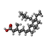  Mass: 354.526 Da / Num. of mol.: 1 / Source method: obtained synthetically / Formula: C24H34O2 Mass: 354.526 Da / Num. of mol.: 1 / Source method: obtained synthetically / Formula: C24H34O2 |
| #4: Water | ChemComp-HOH /  Mass: 18.015 Da / Num. of mol.: 124 / Source method: isolated from a natural source / Formula: H2O Mass: 18.015 Da / Num. of mol.: 124 / Source method: isolated from a natural source / Formula: H2O |
-Experimental details
-Experiment
| Experiment | Method: X-RAY DIFFRACTION / Number of used crystals: 1 |
|---|
- Sample preparation
| Crystal | Density Matthews: 2.22 Å3/Da / Density % sol: 44.7 % |
|---|---|
| Crystal grow | Temperature: 295 K / Method: vapor diffusion, hanging drop / pH: 7 Details: 5-15% PEG4000, 4-12% Glycerol, 0.1M Bis-Tris pH=7.0, VAPOR DIFFUSION, HANGING DROP, temperature 295K |
-Data collection
| Diffraction | Mean temperature: 100 K | ||||||||||||
|---|---|---|---|---|---|---|---|---|---|---|---|---|---|
| Diffraction source | Source: SYNCHROTRON / Site: APS  / Beamline: 22-BM / Wavelength: 0.97623 Å / Beamline: 22-BM / Wavelength: 0.97623 Å | ||||||||||||
| Detector | Type: MAR CCD 165 mm / Detector: CCD / Date: Feb 17, 2005 | ||||||||||||
| Radiation | Monochromator: double crystal - liqued nitrogen cooled / Protocol: SINGLE WAVELENGTH / Monochromatic (M) / Laue (L): M / Scattering type: x-ray | ||||||||||||
| Radiation wavelength | Wavelength: 0.97623 Å / Relative weight: 1 | ||||||||||||
| Reflection | Resolution: 1.9→20 Å / Num. obs: 238303 / % possible obs: 99.9 % / Observed criterion σ(F): 2 / Observed criterion σ(I): 2 / Redundancy: 11.9 % / Rmerge(I) obs: 0.048 / Rsym value: 0.255 | ||||||||||||
| Reflection shell |
|
- Processing
| Software |
| |||||||||||||||||||||||||
|---|---|---|---|---|---|---|---|---|---|---|---|---|---|---|---|---|---|---|---|---|---|---|---|---|---|---|
| Refinement | Method to determine structure: MOLECULAR REPLACEMENT Starting model: 3OAP Resolution: 1.9→20 Å / σ(F): 2 / Stereochemistry target values: Engh & Huber
| |||||||||||||||||||||||||
| Refinement step | Cycle: LAST / Resolution: 1.9→20 Å
| |||||||||||||||||||||||||
| Refine LS restraints |
| |||||||||||||||||||||||||
| LS refinement shell |
|
