 Movie
Movie Controller

[English] 日本語
 Yorodumi
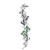
- PDB-4kzd: Crystal structure of an RNA aptamer in complex with fluorophore a... -
+ Open data

- Basic information
| Entry | Database: PDB / ID: 4kzd | |||||||||
|---|---|---|---|---|---|---|---|---|---|---|
| Title | Crystal structure of an RNA aptamer in complex with fluorophore and Fab | |||||||||
 Components Components |
| |||||||||
 Keywords Keywords | immune system/rna / G-quadruplex / Fluorescence / Fluorophore binding / In vitro / immune system-rna complex | |||||||||
| Function / homology | Immunoglobulins / Immunoglobulin-like / Sandwich / Mainly Beta / sucrose / Chem-1TU / : / RNA / RNA (> 10) Function and homology information Function and homology information | |||||||||
| Biological species |  | |||||||||
| Method |  X-RAY DIFFRACTION / SYNCHROTRON / MOLECULAR REPLACEMENT / Resolution: 2.186 Å X-RAY DIFFRACTION / SYNCHROTRON / MOLECULAR REPLACEMENT / Resolution: 2.186 Å | |||||||||
 Authors Authors | Huang, H. / Suslov, N.B. / Li, N. / Koldobskaya, Y. / Rice, P.A. / Piccirilli, J.A. | |||||||||
 Citation Citation | Journal: Nat.Chem.Biol. / Year: 2014 Title: A G-quadruplex-containing RNA activates fluorescence in a GFP-like fluorophore. Authors: Huang, H. / Suslov, N.B. / Li, N.S. / Shelke, S.A. / Evans, M.E. / Koldobskaya, Y. / Rice, P.A. / Piccirilli, J.A. | |||||||||
| History |
|
- Structure visualization
| Structure viewer | Molecule: MolmilJmol/JSmol |
|---|
- Downloads & links
-Download
| PDBx/mmCIF format | 4kzd.cif.gz | 162.7 KB | Display | PDBx/mmCIF format |
|---|---|---|---|---|
| PDB format | pdb4kzd.ent.gz | 119 KB | Display | PDB format |
| PDBx/mmJSON format | 4kzd.json.gz | Tree view | PDBx/mmJSON format | |
| Others |  Other downloads Other downloads |
-Validation report
| Arichive directory | https://data.pdbj.org/pub/pdb/validation_reports/kz/4kzdftp://data.pdbj.org/pub/pdb/validation_reports/kz/4kzd | HTTPS FTP |
|---|
-Related structure data
| Related structure data |  4kzeC  4q9qC  4q9rC  3ivkS S: Starting model for refinement C: citing same article ( |
|---|---|
| Similar structure data |

-Links
 PDBj
PDBj


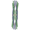
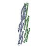
- Assembly
| Deposited unit | 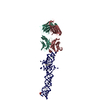
| ||||||||
|---|---|---|---|---|---|---|---|---|---|
| 1 |
| ||||||||
| 2 | 
| ||||||||
| 3 | 
| ||||||||
| 4 | 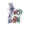
| ||||||||
| Unit cell |
|
-Components
-Antibody , 2 types, 2 molecules HL
| #1: Antibody | Mass: 24665.510 Da / Num. of mol.: 1 Source method: isolated from a genetically manipulated source Source: (gene. exp.)  |
|---|---|
| #2: Antibody | Mass: 23394.891 Da / Num. of mol.: 1 Source method: isolated from a genetically manipulated source Source: (gene. exp.) |
-RNA chain / Sugars , 2 types, 2 molecules R
| #3: RNA chain | Mass: 27366.256 Da / Num. of mol.: 1 / Source method: obtained synthetically / Details: Spinach RNA aptamer |
|---|---|
| #4: Polysaccharide | beta-D-fructofuranose-(2-1)-alpha-D-glucopyranose / sucrose  Source method: isolated from a genetically manipulated source Details: oligosaccharide with reducing-end-to-reducing-end glycosidic bond References: sucrose |
-Non-polymers , 4 types, 469 molecules 


| #5: Chemical | ChemComp-K /  Mass: 39.098 Da / Num. of mol.: 1 / Source method: obtained synthetically / Formula: K Mass: 39.098 Da / Num. of mol.: 1 / Source method: obtained synthetically / Formula: K |
|---|---|
| #6: Chemical | ChemComp-MG /  Mass: 24.305 Da / Num. of mol.: 1 / Source method: obtained synthetically / Formula: Mg Mass: 24.305 Da / Num. of mol.: 1 / Source method: obtained synthetically / Formula: Mg |
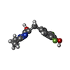
| #7: Chemical | ChemComp-1TU /  Mass: 254.233 Da / Num. of mol.: 1 / Source method: obtained synthetically / Formula: C12H12F2N2O2 Mass: 254.233 Da / Num. of mol.: 1 / Source method: obtained synthetically / Formula: C12H12F2N2O2 |
| #8: Water | ChemComp-HOH / Mass: 18.015 Da / Num. of mol.: 466 / Source method: isolated from a natural source / Formula: H2O |
-Details
| Has protein modification | Y |
|---|
-Experimental details
-Experiment
| Experiment | Method: X-RAY DIFFRACTION / Number of used crystals: 1 |
|---|
- Sample preparation
| Crystal | Density Matthews: 3.47 Å3/Da / Density % sol: 64.51 % |
|---|---|
| Crystal grow | Temperature: 298 K / Method: vapor diffusion, hanging drop / pH: 7 Details: 8% Tacsimate, 20% PEG 3,350, pH 7.0, VAPOR DIFFUSION, HANGING DROP, temperature 298K |
-Data collection
| Diffraction | Mean temperature: 77 K |
|---|---|
| Diffraction source | Source: SYNCHROTRON / Site: APS  / Beamline: 24-ID-C / Wavelength: 0.9795 Å / Beamline: 24-ID-C / Wavelength: 0.9795 Å |
| Detector | Type: ADSC QUANTUM 315 / Detector: CCD / Date: Feb 27, 2013 |
| Radiation | Monochromator: Si(111) / Protocol: SINGLE WAVELENGTH / Monochromatic (M) / Laue (L): M / Scattering type: x-ray |
| Radiation wavelength | Wavelength: 0.9795 Å / Relative weight: 1 |
| Reflection | Resolution: 2.19→46.69 Å / Num. all: 52772 / Num. obs: 52477 / % possible obs: 98.2 % / Observed criterion σ(F): 2 / Observed criterion σ(I): 1 / Redundancy: 3 % / Rmerge(I) obs: 0.059 |
| Reflection shell | Resolution: 2.19→2.3 Å / Redundancy: 2.9 % / Rmerge(I) obs: 0.615 / Mean I/σ(I) obs: 1.8 / % possible all: 98.3 |
- Processing
| Software |
| |||||||||||||||||||||||||||||||||||||||||||||||||||||||||||||||||||||||||||||||||||||||||||||||||||||||||
|---|---|---|---|---|---|---|---|---|---|---|---|---|---|---|---|---|---|---|---|---|---|---|---|---|---|---|---|---|---|---|---|---|---|---|---|---|---|---|---|---|---|---|---|---|---|---|---|---|---|---|---|---|---|---|---|---|---|---|---|---|---|---|---|---|---|---|---|---|---|---|---|---|---|---|---|---|---|---|---|---|---|---|---|---|---|---|---|---|---|---|---|---|---|---|---|---|---|---|---|---|---|---|---|---|---|---|
| Refinement | Method to determine structure: MOLECULAR REPLACEMENT Starting model: PDB entry 3IVK Resolution: 2.186→46.687 Å / SU ML: 0.29 / σ(F): 1.34 / Phase error: 23.1 / Stereochemistry target values: ML
| |||||||||||||||||||||||||||||||||||||||||||||||||||||||||||||||||||||||||||||||||||||||||||||||||||||||||
| Solvent computation | Shrinkage radii: 0.9 Å / VDW probe radii: 1.11 Å / Solvent model: FLAT BULK SOLVENT MODEL | |||||||||||||||||||||||||||||||||||||||||||||||||||||||||||||||||||||||||||||||||||||||||||||||||||||||||
| Refinement step | Cycle: LAST / Resolution: 2.186→46.687 Å
| |||||||||||||||||||||||||||||||||||||||||||||||||||||||||||||||||||||||||||||||||||||||||||||||||||||||||
| Refine LS restraints |
| |||||||||||||||||||||||||||||||||||||||||||||||||||||||||||||||||||||||||||||||||||||||||||||||||||||||||
| LS refinement shell |
|
