 Movie
Movie Controller

[English] 日本語
 Yorodumi
- EMDB-44971: Sub-tomogram average of two pairs of RSV F trimers from the surfa... -
+ Open data

- Basic information
| Entry |  | ||||||||||||
|---|---|---|---|---|---|---|---|---|---|---|---|---|---|
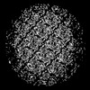
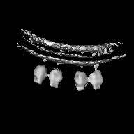
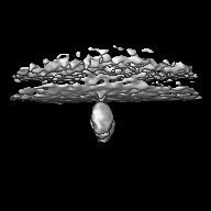
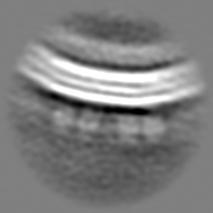
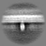
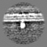
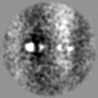
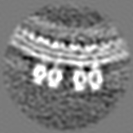
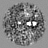
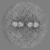
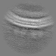
| Title | Sub-tomogram average of two pairs of RSV F trimers from the surface of native virions released from RSV-infected BEAS-2B cells cultured on EM grids | ||||||||||||
 Map data Map data | |||||||||||||
 Sample Sample |
| ||||||||||||
 Keywords Keywords | Membrane Fusion / VIRAL PROTEIN | ||||||||||||
| Biological species |  Human respiratory syncytial virus A2 Human respiratory syncytial virus A2 | ||||||||||||
| Method | subtomogram averaging / cryo EM / Resolution: 36.0 Å | ||||||||||||
 Authors Authors | Sibert BS / Wright ER | ||||||||||||
| Funding support |  United States, 3 items United States, 3 items
| ||||||||||||
 Citation Citation | Journal: Nat Commun / Year: 2024 Title: Assembly of respiratory syncytial virus matrix protein lattice and its coordination with fusion glycoprotein trimers. Authors: Bryan S Sibert / Joseph Y Kim / Jie E Yang / Zunlong Ke / Christopher C Stobart / Martin L Moore / Elizabeth R Wright / Abstract: Respiratory syncytial virus (RSV) is an enveloped, filamentous, negative-strand RNA virus that causes significant respiratory illness worldwide. RSV vaccines are available, however there is still ...Respiratory syncytial virus (RSV) is an enveloped, filamentous, negative-strand RNA virus that causes significant respiratory illness worldwide. RSV vaccines are available, however there is still significant need for research to support the development of vaccines and therapeutics against RSV and related Mononegavirales viruses. Individual virions vary in size, with an average diameter of ~130 nm and ranging from ~500 nm to over 10 µm in length. Though the general arrangement of structural proteins in virions is known, we use cryo-electron tomography and sub-tomogram averaging to determine the molecular organization of RSV structural proteins. We show that the peripheral membrane-associated RSV matrix (M) protein is arranged in a packed helical-like lattice of M-dimers. We report that RSV F glycoprotein is frequently observed as pairs of trimers oriented in an anti-parallel conformation to support potential interactions between trimers. Our sub-tomogram averages indicate the positioning of F-trimer pairs is correlated with the underlying M lattice. These results provide insight into RSV virion organization and may aid in the development of RSV vaccines and anti-viral targets. | ||||||||||||
| History |
|
- Structure visualization
| Supplemental images |
|---|

- Downloads & links
-EMDB archive
| Map data | emd_44971.map.gz | 25.2 MB |  EMDB map data format EMDB map data format | |
|---|---|---|---|---|
| Header (meta data) | emd-44971-v30.xmlemd-44971.xml | 15.6 KB 15.6 KB | Display Display | EMDB header |
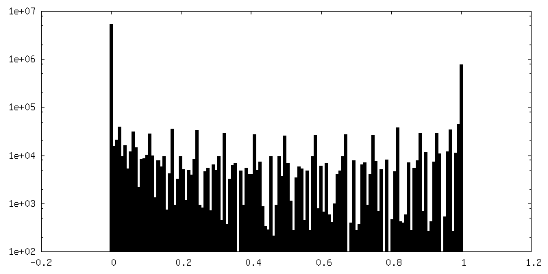
| FSC (resolution estimation) | emd_44971_fsc.xml | 7 KB | Display | FSC data file |
| Images |  emd_44971.png emd_44971.png | 71.2 KB | ||
| Masks | emd_44971_msk_1.map | 27 MB | Mask map | |
| Filedesc metadata | emd-44971.cif.gz | 4.8 KB | ||
| Others | emd_44971_half_map_1.map.gzemd_44971_half_map_2.map.gz | 13.4 MB 13.4 MB | ||
| Archive directory |  http://ftp.pdbj.org/pub/emdb/structures/EMD-44971ftp://ftp.pdbj.org/pub/emdb/structures/EMD-44971 http://ftp.pdbj.org/pub/emdb/structures/EMD-44971ftp://ftp.pdbj.org/pub/emdb/structures/EMD-44971 | HTTPS FTP |
-Validation report
| Summary document | emd_44971_validation.pdf.gz | 1 MB | Display | EMDB validaton report |
|---|---|---|---|---|
| Full document | emd_44971_full_validation.pdf.gz | 1 MB | Display | |
| Data in XML | emd_44971_validation.xml.gz | 12.5 KB | Display | |
| Data in CIF | emd_44971_validation.cif.gz | 17.1 KB | Display | |
| Arichive directory | https://ftp.pdbj.org/pub/emdb/validation_reports/EMD-44971ftp://ftp.pdbj.org/pub/emdb/validation_reports/EMD-44971 | HTTPS FTP |
-Related structure data


-Links
| EMDB pages | EMDB (EBI/PDBe) / EMDataResource |
|---|
-Map
| File | Download / File: emd_44971.map.gz / Format: CCP4 / Size: 27 MB / Type: IMAGE STORED AS FLOATING POINT NUMBER (4 BYTES) | ||||||||||||||||||||||||||||||||||||
|---|---|---|---|---|---|---|---|---|---|---|---|---|---|---|---|---|---|---|---|---|---|---|---|---|---|---|---|---|---|---|---|---|---|---|---|---|---|
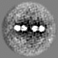
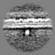
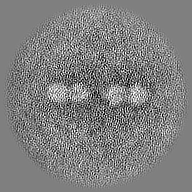
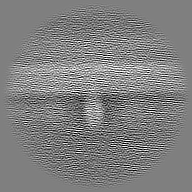
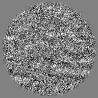
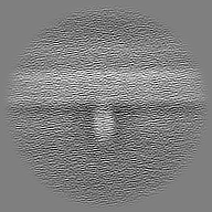
| Projections & slices | Image control
Images are generated by Spider. | ||||||||||||||||||||||||||||||||||||
| Voxel size | X=Y=Z: 3.386 Å | ||||||||||||||||||||||||||||||||||||
| Density |
| ||||||||||||||||||||||||||||||||||||
| Symmetry | Space group: 1 | ||||||||||||||||||||||||||||||||||||
| Details | EMDB XML:
|
 Z (Sec.)
 Y (Row.)
 X (Col.)


-Supplemental data
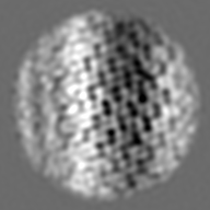
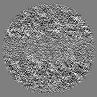
-Mask #1
| File | emd_44971_msk_1.map | ||||||||||||
|---|---|---|---|---|---|---|---|---|---|---|---|---|---|
| Projections & Slices |
| ||||||||||||
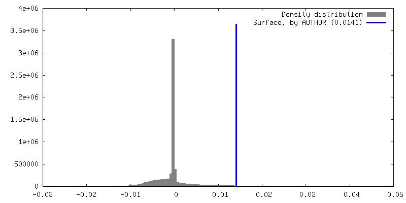
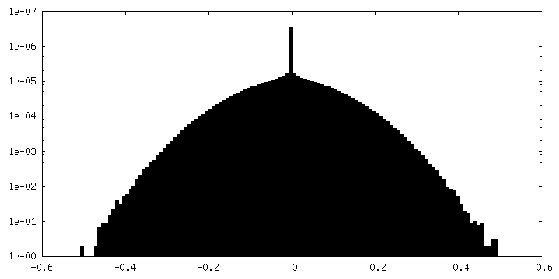
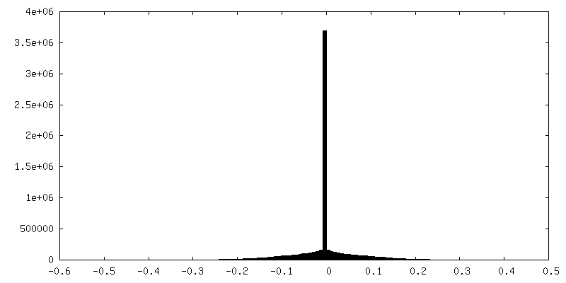
| Density Histograms |


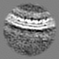
-Half map: #1
| File | emd_44971_half_map_1.map | ||||||||||||
|---|---|---|---|---|---|---|---|---|---|---|---|---|---|
| Projections & Slices |
| ||||||||||||
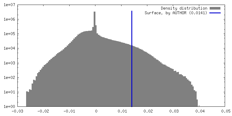
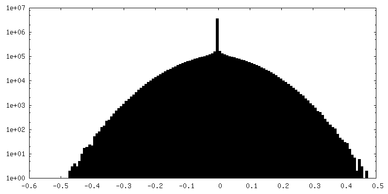
| Density Histograms |


-Half map: #2
| File | emd_44971_half_map_2.map | ||||||||||||
|---|---|---|---|---|---|---|---|---|---|---|---|---|---|
| Projections & Slices |
| ||||||||||||
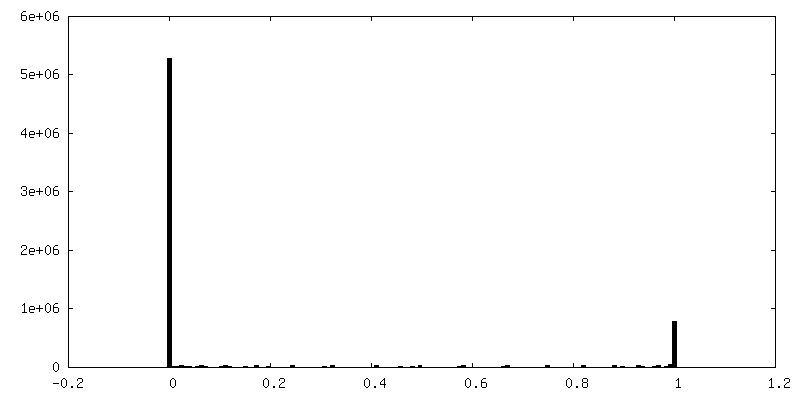
| Density Histograms |


- Sample components
-Entire : Human respiratory syncytial virus A2
| Entire | Name: Human respiratory syncytial virus A2 |
|---|---|
| Components |
|
-Supramolecule #1: Human respiratory syncytial virus A2
| Supramolecule | Name: Human respiratory syncytial virus A2 / type: virus / ID: 1 / Parent: 0 Details: The strain was kindly provided by Dr. Martin L. Moore. The reference is PMCID: PMC3492879/PMID:23062737 NCBI-ID: 11259 / Sci species name: Human respiratory syncytial virus A2 / Sci species strain: rA2-mK+ / Virus type: VIRION / Virus isolate: STRAIN / Virus enveloped: Yes / Virus empty: No |
|---|---|
| Host (natural) | Organism:  Homo sapiens (human) Homo sapiens (human) |
-Experimental details
-Structure determination
| Method | cryo EM |
|---|---|
 Processing Processing | subtomogram averaging |
| Aggregation state | particle |
-Sample preparation
| Buffer | pH: 7.2 / Details: Cells were grown in RPMI medium. |
|---|---|
| Grid | Model: Quantifoil R2/1 / Material: GOLD / Mesh: 200 / Support film - Material: CARBON / Support film - topology: HOLEY ARRAY / Pretreatment - Type: GLOW DISCHARGE / Pretreatment - Time: 60 sec. |
| Vitrification | Cryogen name: ETHANE / Chamber humidity: 80 % / Chamber temperature: 303 K / Instrument: GATAN CRYOPLUNGE 3 |
| Details | Filamentous virions released from BEAS-2B cells grown and infected with RSV A2-mk+ on cryo-TEM grids. |
- Electron microscopy
| Microscope | FEI TITAN KRIOS |
|---|---|
| Specialist optics | Energy filter - Name: GIF Bioquantum / Energy filter - Slit width: 20 eV |
| Image recording | Film or detector model: GATAN K3 BIOQUANTUM (6k x 4k) / Digitization - Dimensions - Width: 5760 pixel / Digitization - Dimensions - Height: 4096 pixel / Average exposure time: 0.35 sec. / Average electron dose: 2.44 e/Å2 |
| Electron beam | Acceleration voltage: 300 kV / Electron source:  FIELD EMISSION GUN FIELD EMISSION GUN |
| Electron optics | C2 aperture diameter: 70.0 µm / Illumination mode: FLOOD BEAM / Imaging mode: BRIGHT FIELD / Cs: 2.7 mm / Nominal defocus max: 6.0 µm / Nominal defocus min: 2.0 µm / Nominal magnification: 53000 |
| Sample stage | Specimen holder model: FEI TITAN KRIOS AUTOGRID HOLDER / Cooling holder cryogen: NITROGEN |
| Experimental equipment |  Model: Titan Krios / Image courtesy: FEI Company |
-Image processing
| Final reconstruction | Applied symmetry - Point group: C1 (asymmetric) / Resolution.type: BY AUTHOR / Resolution: 36.0 Å / Resolution method: FSC 0.5 CUT-OFF / Software - Name: RELION (ver. 4.0) / Number subtomograms used: 1132 |
|---|---|
| Extraction | Number tomograms: 27 / Number images used: 305989 |
| Final angle assignment | Type: MAXIMUM LIKELIHOOD / Software - Name: RELION |
| FSC plot (resolution estimation) |  |
