 Movie
Movie Controller


+ Open data

- Basic information
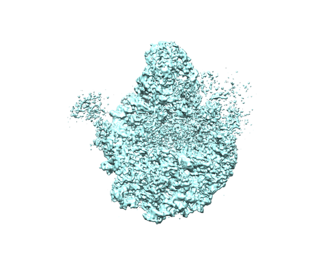
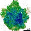
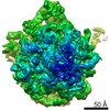
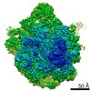
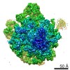
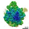
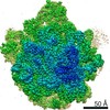
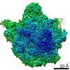
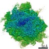
| Entry | Database: EMDB / ID: EMD-4380 | |||||||||
|---|---|---|---|---|---|---|---|---|---|---|
| Title | 50S ribosomal subunit assembly intermediate state 3 | |||||||||
 Map data Map data | ||||||||||
 Sample Sample |
| |||||||||
 Keywords Keywords | assembly precursor 48S 50S biogenesis in vitro reconstitution / RIBOSOME | |||||||||
| Function / homology |  Function and homology information Function and homology informationtranscriptional attenuation / endoribonuclease inhibitor activity / positive regulation of ribosome biogenesis / RNA-binding transcription regulator activity / negative regulation of cytoplasmic translation / DnaA-L2 complex / translation repressor activity / negative regulation of DNA-templated DNA replication initiation / mRNA regulatory element binding translation repressor activity / response to reactive oxygen species ...transcriptional attenuation / endoribonuclease inhibitor activity / positive regulation of ribosome biogenesis / RNA-binding transcription regulator activity / negative regulation of cytoplasmic translation / DnaA-L2 complex / translation repressor activity / negative regulation of DNA-templated DNA replication initiation / mRNA regulatory element binding translation repressor activity / response to reactive oxygen species / cytosolic ribosome assembly / ribosome assembly / assembly of large subunit precursor of preribosome / regulation of cell growth / DNA-templated transcription termination / response to radiation / mRNA 5'-UTR binding / large ribosomal subunit / transferase activity / ribosome binding / 5S rRNA binding / ribosomal large subunit assembly / large ribosomal subunit rRNA binding / cytosolic large ribosomal subunit / cytoplasmic translation / tRNA binding / negative regulation of translation / rRNA binding / structural constituent of ribosome / ribosome / translation / response to antibiotic / negative regulation of DNA-templated transcription / mRNA binding / DNA binding / RNA binding / zinc ion binding / cytoplasm / cytosol Similarity search - Function | |||||||||
| Biological species |  | |||||||||
| Method | single particle reconstruction / cryo EM / Resolution: 4.3 Å | |||||||||
 Authors Authors | Nikolay R / Hilal T | |||||||||
| Funding support |  Germany, 2 items Germany, 2 items
| |||||||||
 Citation Citation | Journal: Mol Cell / Year: 2018 Title: Structural Visualization of the Formation and Activation of the 50S Ribosomal Subunit during In Vitro Reconstitution. Authors: Rainer Nikolay / Tarek Hilal / Bo Qin / Thorsten Mielke / Jörg Bürger / Justus Loerke / Kathrin Textoris-Taube / Knud H Nierhaus / Christian M T Spahn / Abstract: The assembly of ribosomal subunits is an essential prerequisite for protein biosynthesis in all domains of life. Although biochemical and biophysical approaches have advanced our understanding of ...The assembly of ribosomal subunits is an essential prerequisite for protein biosynthesis in all domains of life. Although biochemical and biophysical approaches have advanced our understanding of ribosome assembly, our mechanistic comprehension of this process is still limited. Here, we perform an in vitro reconstitution of the Escherichia coli 50S ribosomal subunit. Late reconstitution products were subjected to high-resolution cryo-electron microscopy and multiparticle refinement analysis to reconstruct five distinct precursors of the 50S subunit with 4.3-3.8 Å resolution. These assembly intermediates define a progressive maturation pathway culminating in a late assembly particle, whose structure is more than 96% identical to a mature 50S subunit. Our structures monitor the formation and stabilization of structural elements in a nascent particle in unprecedented detail and identify the maturation of the rRNA-based peptidyl transferase center as the final critical step along the 50S assembly pathway. | |||||||||
| History |
|
- Structure visualization
| Movie |
Movie viewer |
|---|---|
| Structure viewer | EM map: SurfViewMolmilJmol/JSmol |
| Supplemental images |
- Downloads & links
-EMDB archive
| Map data | emd_4380.map.gz | 70.1 MB | EMDB map data format | |
|---|---|---|---|---|
| Header (meta data) | emd-4380-v30.xmlemd-4380.xml | 37.3 KB 37.3 KB | Display Display | EMDB header |
| Images |  emd_4380.png emd_4380.png | 116.7 KB | ||
| Filedesc metadata | emd-4380.cif.gz | 9 KB | ||
| Archive directory |  http://ftp.pdbj.org/pub/emdb/structures/EMD-4380ftp://ftp.pdbj.org/pub/emdb/structures/EMD-4380 http://ftp.pdbj.org/pub/emdb/structures/EMD-4380ftp://ftp.pdbj.org/pub/emdb/structures/EMD-4380 | HTTPS FTP |
-Related structure data
| Related structure data | 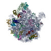 6gc4MC 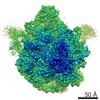 4378C 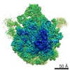 4379C 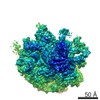 4381C 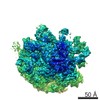 4382C 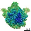 4383C  6gbzC  6gc0C  6gc6C  6gc7C  6gc8C C: citing same article ( M: atomic model generated by this map |
|---|---|
| Similar structure data |
-Links
| EMDB pages | EMDB (EBI/PDBe) / EMDataResource |
|---|---|
| Related items in Molecule of the Month |


-Map
| File | Download / File: emd_4380.map.gz / Format: CCP4 / Size: 75.1 MB / Type: IMAGE STORED AS FLOATING POINT NUMBER (4 BYTES) | ||||||||||||||||||||||||||||||||||||||||||||||||||||||||||||
|---|---|---|---|---|---|---|---|---|---|---|---|---|---|---|---|---|---|---|---|---|---|---|---|---|---|---|---|---|---|---|---|---|---|---|---|---|---|---|---|---|---|---|---|---|---|---|---|---|---|---|---|---|---|---|---|---|---|---|---|---|---|
| Projections & slices | Image control
Images are generated by Spider. | ||||||||||||||||||||||||||||||||||||||||||||||||||||||||||||
| Voxel size | X=Y=Z: 1.24 Å | ||||||||||||||||||||||||||||||||||||||||||||||||||||||||||||
| Density |
| ||||||||||||||||||||||||||||||||||||||||||||||||||||||||||||
| Symmetry | Space group: 1 | ||||||||||||||||||||||||||||||||||||||||||||||||||||||||||||
| Details | EMDB XML:
CCP4 map header:
| ||||||||||||||||||||||||||||||||||||||||||||||||||||||||||||
 Z (Sec.)
 Y (Row.)
 X (Col.)


-Supplemental data
- Sample components
+Entire : 50S ribosomal assembly intermediate state 3
+Supramolecule #1: 50S ribosomal assembly intermediate state 3
+Macromolecule #1: 50S ribosomal protein L25
+Macromolecule #3: 50S ribosomal protein L2
+Macromolecule #4: 50S ribosomal protein L3
+Macromolecule #5: 50S ribosomal protein L4
+Macromolecule #6: 50S ribosomal protein L6
+Macromolecule #7: 50S ribosomal protein L13
+Macromolecule #8: 50S ribosomal protein L14
+Macromolecule #9: 50S ribosomal protein L15
+Macromolecule #10: 50S ribosomal protein L17
+Macromolecule #11: 50S ribosomal protein L19
+Macromolecule #12: 50S ribosomal protein L20
+Macromolecule #13: 50S ribosomal protein L21
+Macromolecule #14: 50S ribosomal protein L22
+Macromolecule #15: 50S ribosomal protein L23
+Macromolecule #16: 50S ribosomal protein L24
+Macromolecule #17: 50S ribosomal protein L28
+Macromolecule #18: 50S ribosomal protein L29
+Macromolecule #19: 50S ribosomal protein L32
+Macromolecule #20: 50S ribosomal protein L34
+Macromolecule #21: 50S ribosomal protein L18
+Macromolecule #22: 50S ribosomal protein L5
+Macromolecule #24: 50S ribosomal protein L30
+Macromolecule #25: 50S ribosomal protein L9
+Macromolecule #26: 50S ribosomal protein L27
+Macromolecule #2: 23S ribosomal RNA
+Macromolecule #23: 5S ribosomal RNA
-Experimental details
-Structure determination
| Method | cryo EM |
|---|---|
 Processing Processing | single particle reconstruction |
| Aggregation state | particle |
-Sample preparation
| Buffer | pH: 7.6 |
|---|---|
| Vitrification | Cryogen name: ETHANE |
- Electron microscopy
| Microscope | FEI POLARA 300 |
|---|---|
| Image recording | Film or detector model: GATAN K2 SUMMIT (4k x 4k) / Detector mode: SUPER-RESOLUTION / Average electron dose: 25.0 e/Å2 |
| Electron beam | Acceleration voltage: 300 kV / Electron source:  FIELD EMISSION GUN FIELD EMISSION GUN |
| Electron optics | Illumination mode: FLOOD BEAM / Imaging mode: BRIGHT FIELD / Nominal magnification: 80645 |
| Experimental equipment |  Model: Tecnai Polara / Image courtesy: FEI Company |
+Image processing
-Atomic model buiding 1
| Refinement | Space: REAL / Protocol: RIGID BODY FIT |
|---|---|
| Output model | PDB-6gc4: |
