 Movie
Movie Controller


+ Open data

- Basic information
| Entry | Database: EMDB / ID: EMD-4093 | |||||||||
|---|---|---|---|---|---|---|---|---|---|---|
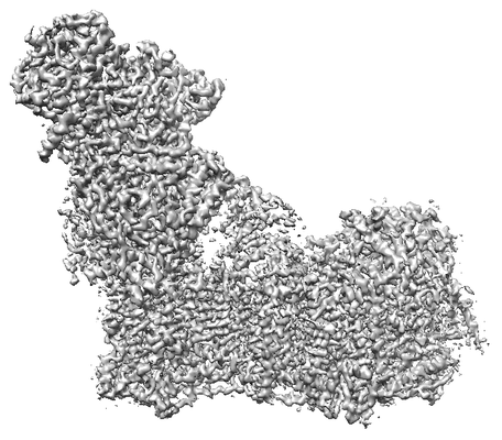
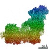
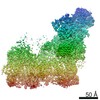
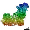
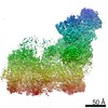
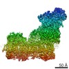
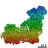
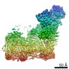
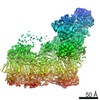
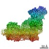
| Title | Entire ovine respiratory complex I, Combined Map | |||||||||
 Map data Map data | ||||||||||
 Sample Sample |
| |||||||||
 Keywords Keywords | NADH:ubiquinone / oxidoreductase / complex I / mammalian / mitochondrial | |||||||||
| Function / homology |  Function and homology information Function and homology informationComplex I biogenesis / : / : / Respiratory electron transport / Mitochondrial protein degradation / NADH:ubiquinone reductase (H+-translocating) / apoptotic mitochondrial changes / ubiquinone binding / mitochondrial ATP synthesis coupled electron transport / mitochondrial electron transport, NADH to ubiquinone ...Complex I biogenesis / : / : / Respiratory electron transport / Mitochondrial protein degradation / NADH:ubiquinone reductase (H+-translocating) / apoptotic mitochondrial changes / ubiquinone binding / mitochondrial ATP synthesis coupled electron transport / mitochondrial electron transport, NADH to ubiquinone / electron transport coupled proton transport / acyl binding / mitochondrial respiratory chain complex I assembly / NADH dehydrogenase activity / oxidoreductase activity, acting on NAD(P)H / membrane => GO:0016020 / respiratory chain complex I / NADH dehydrogenase (ubiquinone) activity / acyl carrier activity / quinone binding / ATP synthesis coupled electron transport / ATP metabolic process / reactive oxygen species metabolic process / aerobic respiration / respiratory electron transport chain / regulation of mitochondrial membrane potential / circadian rhythm / electron transport chain / mitochondrial intermembrane space / 2 iron, 2 sulfur cluster binding / mitochondrial membrane / NAD binding / FMN binding / 4 iron, 4 sulfur cluster binding / response to oxidative stress / oxidoreductase activity / mitochondrial inner membrane / mitochondrial matrix / negative regulation of DNA-templated transcription / apoptotic process / protein-containing complex binding / mitochondrion / nucleoplasm / membrane / metal ion binding / cytoplasm Similarity search - Function | |||||||||
| Biological species |  | |||||||||
| Method | single particle reconstruction / cryo EM / Resolution: 3.9 Å | |||||||||
 Authors Authors | Fiedorczuk K / Letts JA / Sazanov LA | |||||||||
| Funding support |  United Kingdom, 1 items United Kingdom, 1 items
| |||||||||
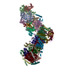
 Citation Citation | Journal: Nature / Year: 2016 Title: Atomic structure of the entire mammalian mitochondrial complex I. Authors: Karol Fiedorczuk / James A Letts / Gianluca Degliesposti / Karol Kaszuba / Mark Skehel / Leonid A Sazanov /  Abstract: Mitochondrial complex I (also known as NADH:ubiquinone oxidoreductase) contributes to cellular energy production by transferring electrons from NADH to ubiquinone coupled to proton translocation ...Mitochondrial complex I (also known as NADH:ubiquinone oxidoreductase) contributes to cellular energy production by transferring electrons from NADH to ubiquinone coupled to proton translocation across the membrane. It is the largest protein assembly of the respiratory chain with a total mass of 970 kilodaltons. Here we present a nearly complete atomic structure of ovine (Ovis aries) mitochondrial complex I at 3.9 Å resolution, solved by cryo-electron microscopy with cross-linking and mass-spectrometry mapping experiments. All 14 conserved core subunits and 31 mitochondria-specific supernumerary subunits are resolved within the L-shaped molecule. The hydrophilic matrix arm comprises flavin mononucleotide and 8 iron-sulfur clusters involved in electron transfer, and the membrane arm contains 78 transmembrane helices, mostly contributed by antiporter-like subunits involved in proton translocation. Supernumerary subunits form an interlinked, stabilizing shell around the conserved core. Tightly bound lipids (including cardiolipins) further stabilize interactions between the hydrophobic subunits. Subunits with possible regulatory roles contain additional cofactors, NADPH and two phosphopantetheine molecules, which are shown to be involved in inter-subunit interactions. We observe two different conformations of the complex, which may be related to the conformationally driven coupling mechanism and to the active-deactive transition of the enzyme. Our structure provides insight into the mechanism, assembly, maturation and dysfunction of mitochondrial complex I, and allows detailed molecular analysis of disease-causing mutations. | |||||||||
| History |
|
- Structure visualization
| Movie |
Movie viewer |
|---|---|
| Structure viewer | EM map: SurfViewMolmilJmol/JSmol |
| Supplemental images |
- Downloads & links
-EMDB archive
| Map data | emd_4093.map.gz | 3 MB | EMDB map data format | |
|---|---|---|---|---|
| Header (meta data) | emd-4093-v30.xmlemd-4093.xml | 71.1 KB 71.1 KB | Display Display | EMDB header |
| Images |  emd_4093.png emd_4093.png | 88.3 KB | ||
| Filedesc metadata | emd-4093.cif.gz | 15.7 KB | ||
| Archive directory |  http://ftp.pdbj.org/pub/emdb/structures/EMD-4093ftp://ftp.pdbj.org/pub/emdb/structures/EMD-4093 http://ftp.pdbj.org/pub/emdb/structures/EMD-4093ftp://ftp.pdbj.org/pub/emdb/structures/EMD-4093 | HTTPS FTP |
-Related structure data
| Related structure data | 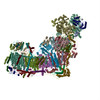 5lnkMC 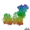 4084C 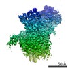 4090C 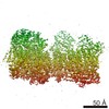 4091C C: citing same article ( M: atomic model generated by this map |
|---|---|
| Similar structure data |

-Links
| EMDB pages | EMDB (EBI/PDBe) / EMDataResource |
|---|---|
| Related items in Molecule of the Month |


-Map
| File | Download / File: emd_4093.map.gz / Format: CCP4 / Size: 14.5 MB / Type: IMAGE STORED AS FLOATING POINT NUMBER (4 BYTES) | ||||||||||||||||||||||||||||||||||||||||||||||||||||||||||||||||||||
|---|---|---|---|---|---|---|---|---|---|---|---|---|---|---|---|---|---|---|---|---|---|---|---|---|---|---|---|---|---|---|---|---|---|---|---|---|---|---|---|---|---|---|---|---|---|---|---|---|---|---|---|---|---|---|---|---|---|---|---|---|---|---|---|---|---|---|---|---|---|
| Projections & slices | Image control
Images are generated by Spider. generated in cubic-lattice coordinate | ||||||||||||||||||||||||||||||||||||||||||||||||||||||||||||||||||||
| Voxel size | X=Y=Z: 1.39 Å | ||||||||||||||||||||||||||||||||||||||||||||||||||||||||||||||||||||
| Density |
| ||||||||||||||||||||||||||||||||||||||||||||||||||||||||||||||||||||
| Symmetry | Space group: 1 | ||||||||||||||||||||||||||||||||||||||||||||||||||||||||||||||||||||
| Details | EMDB XML:
CCP4 map header:
| ||||||||||||||||||||||||||||||||||||||||||||||||||||||||||||||||||||
 X (Sec.)
 Y (Row.)
 Z (Col.)


-Supplemental data
- Sample components
+Entire : Mitochondrial complex I
+Supramolecule #1: Mitochondrial complex I
+Macromolecule #1: Mitochondrial complex I, 51 kDa subunit
+Macromolecule #2: Mitochondrial complex I, 24 kDa subunit
+Macromolecule #3: Mitochondrial complex I, 75 kDa subunit
+Macromolecule #4: Mitochondrial complex I, 49 kDa subunit
+Macromolecule #5: Mitochondrial complex I, 30 kDa subunit
+Macromolecule #6: Mitochondrial complex I, PSST subunit
+Macromolecule #7: Mitochondrial complex I, TYKY subunit
+Macromolecule #8: Mitochondrial complex I, ND1 subunit
+Macromolecule #9: Mitochondrial complex I, ND2 subunit
+Macromolecule #10: Mitochondrial complex I, ND3 subunit
+Macromolecule #11: Mitochondrial complex I, ND4 subunit
+Macromolecule #12: Mitochondrial complex I, ND4L subunit
+Macromolecule #13: Mitochondrial complex I, ND5 subunit
+Macromolecule #14: Mitochondrial complex I, ND6 subunit
+Macromolecule #15: Mitochondrial complex I, 10 kDa subunit
+Macromolecule #16: Mitochondrial complex I, 13 kDa subunit
+Macromolecule #17: Mitochondrial complex I, 18 kDa subunit
+Macromolecule #18: Mitochondrial complex I, 39 kDa subunit
+Macromolecule #19: Mitochondrial complex I, B8 subunit
+Macromolecule #20: Mitochondrial complex I, B13 subunit
+Macromolecule #21: Mitochondrial complex I, B14 subunit
+Macromolecule #22: Mitochondrial complex I, B14.5a subunit
+Macromolecule #23: Mitochondrial complex I, B17.2 subunit
+Macromolecule #24: Acyl carrier protein
+Macromolecule #25: Mitochondrial complex I, 42 kDa subunit
+Macromolecule #26: Mitochondrial complex I, 15 kDa subunit
+Macromolecule #27: Mitochondrial complex I, B9 subunit
+Macromolecule #28: Mitochondrial complex I, B12 subunit
+Macromolecule #29: Mitochondrial complex I, B14.5b subunit
+Macromolecule #30: Mitochondrial complex I, B15 subunit
+Macromolecule #31: Mitochondrial complex I, B16.6 subunit
+Macromolecule #32: Mitochondrial complex I, B17 subunit
+Macromolecule #33: Mitochondrial complex I, B18 subunit
+Macromolecule #34: Mitochondrial complex I, B22 subunit
+Macromolecule #35: Mitochondrial complex I, AGGG subunit
+Macromolecule #36: NADH dehydrogenase [ubiquinone] 1 beta subcomplex subunit 8, mito...
+Macromolecule #37: Mitochondrial complex I, ESSS subunit
+Macromolecule #38: Mitochondrial complex I, KFYI subunit
+Macromolecule #39: Mitochondrial complex I, MNLL subunit
+Macromolecule #40: Mitochondrial complex I, MWFE subunit
+Macromolecule #41: Mitochondrial complex I, PDSW subunit
+Macromolecule #42: Mitochondrial complex I, PGIV subunit
+Macromolecule #43: Mitochondrial complex I, SGDH subunit
+Macromolecule #44: Mitochondrial complex I, B14.7 subunit
+Macromolecule #45: IRON/SULFUR CLUSTER

+Macromolecule #46: FLAVIN MONONUCLEOTIDE

+Macromolecule #47: FE2/S2 (INORGANIC) CLUSTER

+Macromolecule #48: 1,2-Distearoyl-sn-glycerophosphoethanolamine

+Macromolecule #49: 1,2-DIACYL-SN-GLYCERO-3-PHOSPHOCHOLINE

+Macromolecule #50: CARDIOLIPIN

+Macromolecule #51: ZINC ION
+Macromolecule #52: NADPH DIHYDRO-NICOTINAMIDE-ADENINE-DINUCLEOTIDE PHOSPHATE

+Macromolecule #53: S-[2-({N-[(2S)-2-hydroxy-3,3-dimethyl-4-(phosphonooxy)butanoyl]-b...
+Macromolecule #54: 4'-PHOSPHOPANTETHEINE
-Experimental details
-Structure determination
| Method | cryo EM |
|---|---|
 Processing Processing | single particle reconstruction |
| Aggregation state | particle |
-Sample preparation
| Concentration | 5 mg/mL | ||||||||||||||||||
|---|---|---|---|---|---|---|---|---|---|---|---|---|---|---|---|---|---|---|---|
| Buffer | pH: 7.4 Component:
| ||||||||||||||||||
| Grid | Model: Quantifoil R0.6/1 / Material: COPPER / Mesh: 300 / Support film - Material: CARBON / Support film - topology: HOLEY / Pretreatment - Type: GLOW DISCHARGE / Pretreatment - Time: 120 sec. / Pretreatment - Atmosphere: AIR | ||||||||||||||||||
| Vitrification | Cryogen name: ETHANE / Chamber humidity: 90 % / Chamber temperature: 277 K / Instrument: FEI VITROBOT MARK IV / Details: 34s blotting. |
- Electron microscopy
| Microscope | FEI TITAN KRIOS |
|---|---|
| Image recording | Film or detector model: FEI FALCON II (4k x 4k) / Detector mode: INTEGRATING / Digitization - Dimensions - Width: 4096 pixel / Digitization - Dimensions - Height: 4096 pixel / Digitization - Frames/image: 1-7 / Number grids imaged: 2 / Number real images: 2600 / Average exposure time: 1.0 sec. / Average electron dose: 26.0 e/Å2 |
| Electron beam | Acceleration voltage: 300 kV / Electron source:  FIELD EMISSION GUN FIELD EMISSION GUN |
| Electron optics | Calibrated magnification: 100720 / Illumination mode: FLOOD BEAM / Imaging mode: BRIGHT FIELD / Cs: 2.7 mm / Nominal defocus max: 3.5 µm / Nominal defocus min: 0.5 µm / Nominal magnification: 59000 |
| Sample stage | Specimen holder model: FEI TITAN KRIOS AUTOGRID HOLDER / Cooling holder cryogen: NITROGEN |
| Experimental equipment |  Model: Titan Krios / Image courtesy: FEI Company |
+Image processing
-Atomic model buiding 1
| Initial model | PDB ID: Chain - Source name: PDB / Chain - Initial model type: experimental model Details: For the fourteen core subunits we used as a starting point our previous structure from T. thermophilus (PDB 4HEA) with sequence adjusted to ovine and side-chains rebuilt in SQWRL4. These are ...Details: For the fourteen core subunits we used as a starting point our previous structure from T. thermophilus (PDB 4HEA) with sequence adjusted to ovine and side-chains rebuilt in SQWRL4. These are chains 1, 2, 3, 4, 5, 6, 9, H, A, J, K, N, M and L (with the same chain IDs as in 4HEA). For the remaining supernumerary subunits it was mostly de novo building with some help from homology modeling, which in most cases was not very useful due to low sequence homology. In few cases models produced by Phyre2 server were useful and were used as a starting point, due to the presence of templates with high coverage and confidence: 42kDa (chain k) - PDB ID 1P6X (xray) 39kDa (chain d) - PDB ID 3WJ7 (xray) SDAPs (chains j and X) - PDB ID 2DNW (NMR) B8 (chain e) - PDB ID 1S3A (NMR) |
|---|---|
| Refinement | Space: REAL / Protocol: AB INITIO MODEL |
| Output model | PDB-5lnk: |

