 Movie
Movie Controller


+ Open data

- Basic information
| Entry | Database: EMDB / ID: EMD-4089 | |||||||||
|---|---|---|---|---|---|---|---|---|---|---|
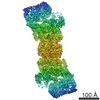
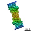
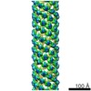
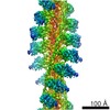
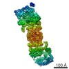
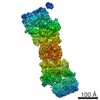
| Title | The human 26S Proteasome at 6.8 Ang. | |||||||||
 Map data Map data | ||||||||||
 Sample Sample |
| |||||||||
 Keywords Keywords | 26S Proteasome / Cryo-EM / Single Particle Analysis / Homology Modelling / hydrolase | |||||||||
| Function / homology |  Function and homology information Function and homology informationpositive regulation of inclusion body assembly / thyrotropin-releasing hormone receptor binding / host-mediated perturbation of viral transcription / Impaired BRCA2 translocation to the nucleus / Impaired BRCA2 binding to SEM1 (DSS1) / cytosolic proteasome complex / Hydrolases; Acting on peptide bonds (peptidases); Omega peptidases / proteasome accessory complex / integrator complex / purine ribonucleoside triphosphate binding ...positive regulation of inclusion body assembly / thyrotropin-releasing hormone receptor binding / host-mediated perturbation of viral transcription / Impaired BRCA2 translocation to the nucleus / Impaired BRCA2 binding to SEM1 (DSS1) / cytosolic proteasome complex / Hydrolases; Acting on peptide bonds (peptidases); Omega peptidases / proteasome accessory complex / integrator complex / purine ribonucleoside triphosphate binding / meiosis I / proteasome regulatory particle / positive regulation of proteasomal protein catabolic process / proteasome-activating activity / proteasome regulatory particle, lid subcomplex / proteasome regulatory particle, base subcomplex / metal-dependent deubiquitinase activity / negative regulation of programmed cell death / protein K63-linked deubiquitination / Regulation of ornithine decarboxylase (ODC) / Proteasome assembly / Homologous DNA Pairing and Strand Exchange / Defective homologous recombination repair (HRR) due to BRCA1 loss of function / Defective HDR through Homologous Recombination Repair (HRR) due to PALB2 loss of BRCA1 binding function / Defective HDR through Homologous Recombination Repair (HRR) due to PALB2 loss of BRCA2/RAD51/RAD51C binding function / Cross-presentation of soluble exogenous antigens (endosomes) / Resolution of D-loop Structures through Synthesis-Dependent Strand Annealing (SDSA) / proteasome core complex / Resolution of D-loop Structures through Holliday Junction Intermediates / Somitogenesis / K63-linked deubiquitinase activity / Impaired BRCA2 binding to RAD51 / proteasome binding / transcription factor binding / regulation of protein catabolic process / myofibril / proteasome storage granule / Presynaptic phase of homologous DNA pairing and strand exchange / general transcription initiation factor binding / blastocyst development / polyubiquitin modification-dependent protein binding / positive regulation of RNA polymerase II transcription preinitiation complex assembly / immune system process / protein deubiquitination / endopeptidase activator activity / NF-kappaB binding / proteasome endopeptidase complex / proteasome core complex, beta-subunit complex / proteasome assembly / threonine-type endopeptidase activity / proteasome core complex, alpha-subunit complex / mRNA export from nucleus / SARS-CoV-1 targets host intracellular signalling and regulatory pathways / inclusion body / enzyme regulator activity / ERAD pathway / regulation of proteasomal protein catabolic process / proteasome complex / proteolysis involved in protein catabolic process / sarcomere / Regulation of activated PAK-2p34 by proteasome mediated degradation / Autodegradation of Cdh1 by Cdh1:APC/C / APC/C:Cdc20 mediated degradation of Securin / N-glycan trimming in the ER and Calnexin/Calreticulin cycle / Asymmetric localization of PCP proteins / Ubiquitin-dependent degradation of Cyclin D / SCF-beta-TrCP mediated degradation of Emi1 / NIK-->noncanonical NF-kB signaling / stem cell differentiation / TNFR2 non-canonical NF-kB pathway / AUF1 (hnRNP D0) binds and destabilizes mRNA / Vpu mediated degradation of CD4 / Assembly of the pre-replicative complex / Ubiquitin-Mediated Degradation of Phosphorylated Cdc25A / Degradation of DVL / Cdc20:Phospho-APC/C mediated degradation of Cyclin A / Dectin-1 mediated noncanonical NF-kB signaling / lipopolysaccharide binding / Degradation of AXIN / Hh mutants are degraded by ERAD / negative regulation of inflammatory response to antigenic stimulus / Activation of NF-kappaB in B cells / P-body / Degradation of GLI1 by the proteasome / Hedgehog ligand biogenesis / G2/M Checkpoints / Defective CFTR causes cystic fibrosis / GSK3B and BTRC:CUL1-mediated-degradation of NFE2L2 / Autodegradation of the E3 ubiquitin ligase COP1 / Negative regulation of NOTCH4 signaling / Vif-mediated degradation of APOBEC3G / Regulation of RUNX3 expression and activity / Hedgehog 'on' state / double-strand break repair via homologous recombination / Degradation of GLI2 by the proteasome / GLI3 is processed to GLI3R by the proteasome / FBXL7 down-regulates AURKA during mitotic entry and in early mitosis / APC/C:Cdh1 mediated degradation of Cdc20 and other APC/C:Cdh1 targeted proteins in late mitosis/early G1 / : / MAPK6/MAPK4 signaling Similarity search - Function | |||||||||
| Biological species |  Homo sapiens (human) Homo sapiens (human) | |||||||||
| Method | single particle reconstruction / cryo EM / Resolution: 6.8 Å | |||||||||
 Authors Authors | Schweitzer A / Beck F | |||||||||
 Citation Citation | Journal: Mol Cell Proteomics / Year: 2017 Title: Molecular Details Underlying Dynamic Structures and Regulation of the Human 26S Proteasome. Authors: Xiaorong Wang / Peter Cimermancic / Clinton Yu / Andreas Schweitzer / Nikita Chopra / James L Engel / Charles Greenberg / Alexander S Huszagh / Florian Beck / Eri Sakata / Yingying Yang / ...Authors: Xiaorong Wang / Peter Cimermancic / Clinton Yu / Andreas Schweitzer / Nikita Chopra / James L Engel / Charles Greenberg / Alexander S Huszagh / Florian Beck / Eri Sakata / Yingying Yang / Eric J Novitsky / Alexander Leitner / Paolo Nanni / Abdullah Kahraman / Xing Guo / Jack E Dixon / Scott D Rychnovsky / Ruedi Aebersold / Wolfgang Baumeister / Andrej Sali / Lan Huang /    Abstract: The 26S proteasome is the macromolecular machine responsible for ATP/ubiquitin dependent degradation. As aberration in proteasomal degradation has been implicated in many human diseases, structural ...The 26S proteasome is the macromolecular machine responsible for ATP/ubiquitin dependent degradation. As aberration in proteasomal degradation has been implicated in many human diseases, structural analysis of the human 26S proteasome complex is essential to advance our understanding of its action and regulation mechanisms. In recent years, cross-linking mass spectrometry (XL-MS) has emerged as a powerful tool for elucidating structural topologies of large protein assemblies, with its unique capability of studying protein complexes in cells. To facilitate the identification of cross-linked peptides, we have previously developed a robust amine reactive sulfoxide-containing MS-cleavable cross-linker, disuccinimidyl sulfoxide (DSSO). To better understand the structure and regulation of the human 26S proteasome, we have established new DSSO-based and XL-MS workflows by coupling with HB-tag based affinity purification to comprehensively examine protein-protein interactions within the 26S proteasome. In total, we have identified 447 unique lysine-to-lysine linkages delineating 67 interprotein and 26 intraprotein interactions, representing the largest cross-link dataset for proteasome complexes. In combination with EM maps and computational modeling, the architecture of the 26S proteasome was determined to infer its structural dynamics. In particular, three proteasome subunits Rpn1, Rpn6, and Rpt6 displayed multiple conformations that have not been previously reported. Additionally, cross-links between proteasome subunits and 15 proteasome interacting proteins including 9 known and 6 novel ones have been determined to demonstrate their physical interactions at the amino acid level. Our results have provided new insights on the dynamics of the 26S human proteasome and the methodologies presented here can be applied to study other protein complexes. | |||||||||
| History |
|
- Structure visualization
| Movie |
Movie viewer |
|---|---|
| Structure viewer | EM map: SurfViewMolmilJmol/JSmol |
| Supplemental images |

- Downloads & links
-EMDB archive
| Map data | emd_4089.map.gz | 5.3 MB | EMDB map data format | |
|---|---|---|---|---|
| Header (meta data) | emd-4089-v30.xmlemd-4089.xml | 48.9 KB 48.9 KB | Display Display | EMDB header |
| Images | 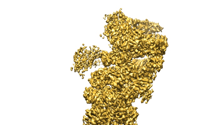 emd_4089.png emd_4089.png | 191.3 KB | ||
| Filedesc metadata | emd-4089.cif.gz | 13.3 KB | ||
| Archive directory |  http://ftp.pdbj.org/pub/emdb/structures/EMD-4089ftp://ftp.pdbj.org/pub/emdb/structures/EMD-4089 http://ftp.pdbj.org/pub/emdb/structures/EMD-4089ftp://ftp.pdbj.org/pub/emdb/structures/EMD-4089 | HTTPS FTP |
-Related structure data
| Related structure data | 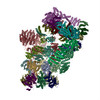 5ln3MC M: atomic model generated by this map C: citing same article ( |
|---|---|
| Similar structure data |
-Links
| EMDB pages | EMDB (EBI/PDBe) / EMDataResource |
|---|---|
| Related items in Molecule of the Month |


-Map
| File | Download / File: emd_4089.map.gz / Format: CCP4 / Size: 83.7 MB / Type: IMAGE STORED AS FLOATING POINT NUMBER (4 BYTES) | ||||||||||||||||||||||||||||||||||||||||||||||||||||||||||||
|---|---|---|---|---|---|---|---|---|---|---|---|---|---|---|---|---|---|---|---|---|---|---|---|---|---|---|---|---|---|---|---|---|---|---|---|---|---|---|---|---|---|---|---|---|---|---|---|---|---|---|---|---|---|---|---|---|---|---|---|---|---|
| Projections & slices | Image control
Images are generated by Spider. | ||||||||||||||||||||||||||||||||||||||||||||||||||||||||||||
| Voxel size | X=Y=Z: 2.16 Å | ||||||||||||||||||||||||||||||||||||||||||||||||||||||||||||
| Density |
| ||||||||||||||||||||||||||||||||||||||||||||||||||||||||||||
| Symmetry | Space group: 1 | ||||||||||||||||||||||||||||||||||||||||||||||||||||||||||||
| Details | EMDB XML:
CCP4 map header:
| ||||||||||||||||||||||||||||||||||||||||||||||||||||||||||||
 Z (Sec.)
 Y (Row.)
 X (Col.)


-Supplemental data
- Sample components
+Entire : Human 26S Proteasome
+Supramolecule #1: Human 26S Proteasome
+Macromolecule #1: 26S proteasome non-ATPase regulatory subunit 2
+Macromolecule #2: Proteasome subunit beta type-6
+Macromolecule #3: Proteasome subunit beta type-7
+Macromolecule #4: Proteasome subunit beta type-3
+Macromolecule #5: Proteasome subunit beta type-2
+Macromolecule #6: Proteasome subunit beta type-5
+Macromolecule #7: Proteasome subunit beta type-1
+Macromolecule #8: Proteasome subunit beta type-4
+Macromolecule #9: Proteasome subunit alpha type-6
+Macromolecule #10: Proteasome subunit alpha type-2
+Macromolecule #11: Proteasome subunit alpha type-4
+Macromolecule #12: Proteasome subunit alpha type-7
+Macromolecule #13: Proteasome subunit alpha type-5
+Macromolecule #14: Proteasome subunit alpha type-1
+Macromolecule #15: Proteasome subunit alpha type-3
+Macromolecule #16: 26S protease regulatory subunit 7
+Macromolecule #17: 26S protease regulatory subunit 4
+Macromolecule #18: 26S protease regulatory subunit 8
+Macromolecule #19: 26S protease regulatory subunit 6B
+Macromolecule #20: 26S protease regulatory subunit 10B
+Macromolecule #21: 26S protease regulatory subunit 6A
+Macromolecule #22: 26S proteasome non-ATPase regulatory subunit 1
+Macromolecule #23: 26S proteasome non-ATPase regulatory subunit 13
+Macromolecule #24: 26S proteasome non-ATPase regulatory subunit 12
+Macromolecule #25: 26S proteasome non-ATPase regulatory subunit 11
+Macromolecule #26: 26S proteasome non-ATPase regulatory subunit 6
+Macromolecule #27: 26S proteasome non-ATPase regulatory subunit 3
+Macromolecule #28: 26S proteasome non-ATPase regulatory subunit 8
+Macromolecule #29: 26S proteasome non-ATPase regulatory subunit 7
+Macromolecule #30: 26S proteasome non-ATPase regulatory subunit 14
+Macromolecule #31: 26S proteasome non-ATPase regulatory subunit 4
+Macromolecule #32: 26S proteasome complex subunit DSS1
-Experimental details
-Structure determination
| Method | cryo EM |
|---|---|
 Processing Processing | single particle reconstruction |
| Aggregation state | particle |
-Sample preparation
| Concentration | 0.5 mg/mL |
|---|---|
| Buffer | pH: 7.5 |
| Sugar embedding | Material: ice |
| Vitrification | Cryogen name: ETHANE |
- Electron microscopy
| Microscope | FEI TITAN KRIOS |
|---|---|
| Image recording | Film or detector model: FEI FALCON II (4k x 4k) / Detector mode: INTEGRATING / Digitization - Dimensions - Width: 4096 pixel / Digitization - Dimensions - Height: 4096 pixel / Digitization - Frames/image: 1-7 / Number grids imaged: 8 / Number real images: 31857 / Average electron dose: 45.0 e/Å2 |
| Electron beam | Acceleration voltage: 300 kV / Electron source:  FIELD EMISSION GUN FIELD EMISSION GUN |
| Electron optics | Illumination mode: FLOOD BEAM / Imaging mode: BRIGHT FIELD / Nominal defocus max: 3.5 µm / Nominal defocus min: 0.8 µm |
| Sample stage | Specimen holder model: FEI TITAN KRIOS AUTOGRID HOLDER / Cooling holder cryogen: NITROGEN |
| Experimental equipment |  Model: Titan Krios / Image courtesy: FEI Company |
-Image processing
| Startup model | Type of model: EMDB MAP EMDB ID: |
|---|---|
| Final reconstruction | Resolution.type: BY AUTHOR / Resolution: 6.8 Å / Resolution method: FSC 0.143 CUT-OFF / Number images used: 252000 |
| Initial angle assignment | Type: PROJECTION MATCHING |
| Final angle assignment | Type: PROJECTION MATCHING |
-Atomic model buiding 1
| Refinement | Space: REAL / Protocol: FLEXIBLE FIT |
|---|---|
| Output model | PDB-5ln3: |
