[English] 日本語
 Yorodumi
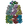
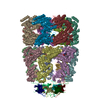
- PDB-3zpz: Visualizing GroEL-ES in the Act of Encapsulating a Non-Native Sub... -
+ Open data
- Basic information
| Entry | Database: PDB / ID: 3zpz | ||||||
|---|---|---|---|---|---|---|---|
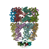
| Title | Visualizing GroEL-ES in the Act of Encapsulating a Non-Native Substrate Protein | ||||||
 Components Components |
| ||||||
 Keywords Keywords | CHAPERONE / PROTEIN FOLDING / HETEROGENEITY | ||||||
| Function / homology |  Function and homology information Function and homology informationGroEL-GroES complex / chaperonin ATPase / virion assembly / : / isomerase activity / protein folding chaperone / ATP-dependent protein folding chaperone / response to radiation / unfolded protein binding / protein folding ...GroEL-GroES complex / chaperonin ATPase / virion assembly / : / isomerase activity / protein folding chaperone / ATP-dependent protein folding chaperone / response to radiation / unfolded protein binding / protein folding / protein-folding chaperone binding / response to heat / protein refolding / magnesium ion binding / ATP hydrolysis activity / ATP binding / metal ion binding / identical protein binding / membrane / cytosol Similarity search - Function | ||||||
| Biological species |   | ||||||
| Method | ELECTRON MICROSCOPY / single particle reconstruction / cryo EM / Resolution: 8.9 Å | ||||||
 Authors Authors | Chen, D.-H. / Madan, D. / Weaver, J. / Lin, Z. / Schroder, G.F. / Chiu, W. / Rye, H.S. | ||||||
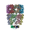
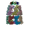
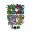
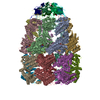
 Citation Citation |  Journal: Cell / Year: 2013 Journal: Cell / Year: 2013Title: Visualizing GroEL/ES in the act of encapsulating a folding protein. Authors: Dong-Hua Chen / Damian Madan / Jeremy Weaver / Zong Lin / Gunnar F Schröder / Wah Chiu / Hays S Rye /  Abstract: The GroEL/ES chaperonin system is required for the assisted folding of many proteins. How these substrate proteins are encapsulated within the GroEL-GroES cavity is poorly understood. Using symmetry- ...The GroEL/ES chaperonin system is required for the assisted folding of many proteins. How these substrate proteins are encapsulated within the GroEL-GroES cavity is poorly understood. Using symmetry-free, single-particle cryo-electron microscopy, we have characterized a chemically modified mutant of GroEL (EL43Py) that is trapped at a normally transient stage of substrate protein encapsulation. We show that the symmetric pattern of the GroEL subunits is broken as the GroEL cis-ring apical domains reorient to accommodate the simultaneous binding of GroES and an incompletely folded substrate protein (RuBisCO). The collapsed RuBisCO folding intermediate binds to the lower segment of two apical domains, as well as to the normally unstructured GroEL C-terminal tails. A comparative structural analysis suggests that the allosteric transitions leading to substrate protein release and folding involve concerted shifts of GroES and the GroEL apical domains and C-terminal tails. | ||||||
| History |
|
- Structure visualization
| Movie |
 Movie viewer Movie viewer |
|---|---|
| Structure viewer | Molecule:  Molmil Molmil Jmol/JSmol Jmol/JSmol |
- Downloads & links
- Download
| PDBx/mmCIF format |  3zpz.cif.gz 3zpz.cif.gz | 1.2 MB | Display |  PDBx/mmCIF format PDBx/mmCIF format |
|---|---|---|---|---|
| PDB format |  pdb3zpz.ent.gz pdb3zpz.ent.gz | 996.9 KB | Display |  PDB format PDB format |
| PDBx/mmJSON format |  3zpz.json.gz 3zpz.json.gz | Tree view |  PDBx/mmJSON format PDBx/mmJSON format | |
| Others |  Other downloads Other downloads |
-Validation report
| Summary document |  3zpz_validation.pdf.gz 3zpz_validation.pdf.gz | 1.4 MB | Display |  wwPDB validaton report wwPDB validaton report |
|---|---|---|---|---|
| Full document |  3zpz_full_validation.pdf.gz 3zpz_full_validation.pdf.gz | 1.6 MB | Display | |
| Data in XML |  3zpz_validation.xml.gz 3zpz_validation.xml.gz | 205.2 KB | Display | |
| Data in CIF |  3zpz_validation.cif.gz 3zpz_validation.cif.gz | 312.2 KB | Display | |
| Arichive directory |  https://data.pdbj.org/pub/pdb/validation_reports/zp/3zpz https://data.pdbj.org/pub/pdb/validation_reports/zp/3zpz ftp://data.pdbj.org/pub/pdb/validation_reports/zp/3zpz ftp://data.pdbj.org/pub/pdb/validation_reports/zp/3zpz | HTTPS FTP |
-Related structure data
| Related structure data |  2325MC  2326C  2327C  3zq0C  3zq1C C: citing same article ( M: map data used to model this data |
|---|---|
| Similar structure data |
- Links
Links
- Assembly
| Deposited unit | 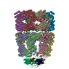
|
|---|---|
| 1 |
|
- Components
| #1: Protein | Mass: 55463.387 Da / Num. of mol.: 14 Source method: isolated from a genetically manipulated source Details: A GROEL VARIANT IN WHICH THE ENDOGENOUS CYS RESIDUES (138,458,519) HAVE BEEN CHANGED TO ALA (GROELCYS0) WAS MODIFIED TO CONTAIN TWO ADDITIONAL MUTATIONS D398A AND S43C. THE D398A MUTATION ...Details: A GROEL VARIANT IN WHICH THE ENDOGENOUS CYS RESIDUES (138,458,519) HAVE BEEN CHANGED TO ALA (GROELCYS0) WAS MODIFIED TO CONTAIN TWO ADDITIONAL MUTATIONS D398A AND S43C. THE D398A MUTATION PREVENTS ATP HYDROLYSIS BY GROEL, WHILE THE S43C MUTATION, LOCATED AT THE TIP OF STEM LOOP AT THE BOTTOM OF THE GROEL CAVITY, PERMITS THE COVALENT ATTACHMENT OF N-1-PYRENE MALEIMIDE TO THIS POSITION. Source: (gene. exp.)   #2: Protein | Mass: 10400.938 Da / Num. of mol.: 7 Source method: isolated from a genetically manipulated source Source: (gene. exp.)   #3: Chemical | ChemComp-MG / #4: Chemical | ChemComp-ADP / |
|---|
-Experimental details
-Experiment
| Experiment | Method: ELECTRON MICROSCOPY |
|---|---|
| EM experiment | Aggregation state: PARTICLE / 3D reconstruction method: single particle reconstruction |
- Sample preparation
| Component | Name: GROEL VARIANT EL43PY CAPPED BY GROES WITH THE ASSISTANCE OF NUCLEOTIDE ATP Type: COMPLEX |
|---|---|
| Buffer solution | Name: 50 MM HEPES, 5 MM KOAC, 10 MM MG(OAC)2, 2 MM DTT / pH: 7.6 / Details: 50 MM HEPES, 5 MM KOAC, 10 MM MG(OAC)2, 2 MM DTT |
| Specimen | Conc.: 6.4 mg/ml / Embedding applied: NO / Shadowing applied: NO / Staining applied: NO / Vitrification applied: YES |
| Specimen support | Details: HOLEY CARBON |
| Vitrification | Instrument: FEI VITROBOT MARK III / Cryogen name: ETHANE Details: VITRIFICATION 1 -- CRYOGEN- ETHANE, HUMIDITY- 95, TEMPERATURE- 98, INSTRUMENT- FEI VITROBOT MARK III, METHOD- BLOT FOR 1 SECOND BEFORE PLUNGING, |
- Electron microscopy imaging
| Microscopy | Model: JEOL KYOTO-3000SFF / Date: Mar 7, 2007 |
|---|---|
| Electron gun | Electron source:  FIELD EMISSION GUN / Accelerating voltage: 300 kV / Illumination mode: FLOOD BEAM FIELD EMISSION GUN / Accelerating voltage: 300 kV / Illumination mode: FLOOD BEAM |
| Electron lens | Mode: OTHER / Nominal magnification: 60000 X / Calibrated magnification: 61060 X / Nominal defocus max: 3500 nm / Nominal defocus min: 1200 nm / Cs: 1.6 mm |
| Specimen holder | Temperature: 11 K |
| Image recording | Electron dose: 36 e/Å2 / Film or detector model: KODAK SO-163 FILM |
| Image scans | Num. digital images: 390 |
- Processing
| EM software |
| ||||||||||||
|---|---|---|---|---|---|---|---|---|---|---|---|---|---|
| CTF correction | Details: EACH MICROGRAPH | ||||||||||||
| Symmetry | Point symmetry: C7 (7 fold cyclic) | ||||||||||||
| 3D reconstruction | Method: FOURIER METHODS / Resolution: 8.9 Å / Num. of particles: 8372 / Nominal pixel size: 2.12 Å / Actual pixel size: 2.08 Å Magnification calibration: THE RMSD BETWEEN ALL EQUATORIAL DOMAINS OF THE FITTED MODELS AND THE X-RAY STRUCTURE OF THE GROEL-GROES-ADP COMPLEX WAS CALCULATED AND THE OPTIMAL PIXEL SIZE WAS CHOSEN AS ...Magnification calibration: THE RMSD BETWEEN ALL EQUATORIAL DOMAINS OF THE FITTED MODELS AND THE X-RAY STRUCTURE OF THE GROEL-GROES-ADP COMPLEX WAS CALCULATED AND THE OPTIMAL PIXEL SIZE WAS CHOSEN AS THE ONE THAT LEADS TO THE SMALLEST RMSD VALUE Details: SUBMISSION BASED ON EXPERIMENTAL DATA FROM EMDB EMD-2325. (DEPOSITION ID: 11454). Symmetry type: POINT | ||||||||||||
| Atomic model building | Protocol: FLEXIBLE FIT / Space: REAL / Target criteria: Cross-correlation coefficient / Details: METHOD--FLEXIBLE REFINEMENT PROTOCOL--X-RAY | ||||||||||||
| Atomic model building | PDB-ID: 1AON Accession code: 1AON / Source name: PDB / Type: experimental model | ||||||||||||
| Refinement | Highest resolution: 8.9 Å | ||||||||||||
| Refinement step | Cycle: LAST / Highest resolution: 8.9 Å
|
 Movie
Movie Controller




 PDBj
PDBj


