[English] 日本語
 Yorodumi
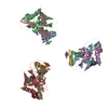
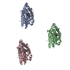
- PDB-3dno: Molecular structure for the HIV-1 gp120 trimer in the CD4-bound state -
+ Open data
- Basic information
| Entry | Database: PDB / ID: 3dno | ||||||
|---|---|---|---|---|---|---|---|
| Title | Molecular structure for the HIV-1 gp120 trimer in the CD4-bound state | ||||||
 Components Components | (HIV-1 envelope glycoprotein gp120) x 3 | ||||||
 Keywords Keywords | VIRAL PROTEIN / HIV-1 / ENVELOPE GLYCOPROTEIN / IMMUNODEFICIENCY VIRUS / gp120 / AIDS / Apoptosis / Cleavage on pair of basic residues / Envelope protein / Fusion protein / Host-virus interaction / Lipoprotein / Membrane / Palmitate / Viral immunoevasion / Virion | ||||||
| Function / homology |  Function and homology information Function and homology informationSynthesis and processing of ENV and VPU / symbiont-mediated evasion of host immune response / positive regulation of establishment of T cell polarity / Alpha-defensins / Dectin-2 family / Binding and entry of HIV virion / positive regulation of plasma membrane raft polarization / positive regulation of receptor clustering / actin filament organization / host cell endosome membrane ...Synthesis and processing of ENV and VPU / symbiont-mediated evasion of host immune response / positive regulation of establishment of T cell polarity / Alpha-defensins / Dectin-2 family / Binding and entry of HIV virion / positive regulation of plasma membrane raft polarization / positive regulation of receptor clustering / actin filament organization / host cell endosome membrane / Assembly Of The HIV Virion / Budding and maturation of HIV virion / clathrin-dependent endocytosis of virus by host cell / viral protein processing / receptor ligand activity / fusion of virus membrane with host plasma membrane / fusion of virus membrane with host endosome membrane / viral envelope / symbiont entry into host cell / virion attachment to host cell / host cell plasma membrane / virion membrane / structural molecule activity / membrane Similarity search - Function | ||||||
| Biological species |  HIV-1 M:B_HXB2R (virus) HIV-1 M:B_HXB2R (virus) | ||||||
| Method | ELECTRON MICROSCOPY / single particle reconstruction / cryo EM / Resolution: 20 Å | ||||||
 Authors Authors | Borgnia, M.J. / Liu, J. / Bartesaghi, A. / Sapiro, G. / Subramaniam, S. | ||||||
 Citation Citation |  Journal: Nature / Year: 2008 Journal: Nature / Year: 2008Title: Molecular architecture of native HIV-1 gp120 trimers. Authors: Jun Liu / Alberto Bartesaghi / Mario J Borgnia / Guillermo Sapiro / Sriram Subramaniam /  Abstract: The envelope glycoproteins (Env) of human and simian immunodeficiency viruses (HIV and SIV, respectively) mediate virus binding to the cell surface receptor CD4 on target cells to initiate infection. ...The envelope glycoproteins (Env) of human and simian immunodeficiency viruses (HIV and SIV, respectively) mediate virus binding to the cell surface receptor CD4 on target cells to initiate infection. Env is a heterodimer of a transmembrane glycoprotein (gp41) and a surface glycoprotein (gp120), and forms trimers on the surface of the viral membrane. Using cryo-electron tomography combined with three-dimensional image classification and averaging, we report the three-dimensional structures of trimeric Env displayed on native HIV-1 in the unliganded state, in complex with the broadly neutralizing antibody b12 and in a ternary complex with CD4 and the 17b antibody. By fitting the known crystal structures of the monomeric gp120 core in the b12- and CD4/17b-bound conformations into the density maps derived by electron tomography, we derive molecular models for the native HIV-1 gp120 trimer in unliganded and CD4-bound states. We demonstrate that CD4 binding results in a major reorganization of the Env trimer, causing an outward rotation and displacement of each gp120 monomer. This appears to be coupled with a rearrangement of the gp41 region along the central axis of the trimer, leading to closer contact between the viral and target cell membranes. Our findings elucidate the structure and conformational changes of trimeric HIV-1 gp120 relevant to antibody neutralization and attachment to target cells. | ||||||
| History |
|
- Structure visualization
| Movie |
 Movie viewer Movie viewer |
|---|---|
| Structure viewer | Molecule:  Molmil Molmil Jmol/JSmol Jmol/JSmol |
- Downloads & links
- Download
| PDBx/mmCIF format |  3dno.cif.gz 3dno.cif.gz | 172.5 KB | Display |  PDBx/mmCIF format PDBx/mmCIF format |
|---|---|---|---|---|
| PDB format |  pdb3dno.ent.gz pdb3dno.ent.gz | 138.4 KB | Display |  PDB format PDB format |
| PDBx/mmJSON format |  3dno.json.gz 3dno.json.gz | Tree view |  PDBx/mmJSON format PDBx/mmJSON format | |
| Others |  Other downloads Other downloads |
-Validation report
| Arichive directory |  https://data.pdbj.org/pub/pdb/validation_reports/dn/3dno https://data.pdbj.org/pub/pdb/validation_reports/dn/3dno ftp://data.pdbj.org/pub/pdb/validation_reports/dn/3dno ftp://data.pdbj.org/pub/pdb/validation_reports/dn/3dno | HTTPS FTP |
|---|
-Related structure data
- Links
Links
- Assembly
| Deposited unit | 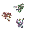
|
|---|---|
| 1 |
|
- Components
| #1: Protein/peptide | Mass: 4225.840 Da / Num. of mol.: 3 / Fragment: Core: Residues 90-124 Source method: isolated from a genetically manipulated source Details: Human immunodeficiency virus type 1 / Source: (gene. exp.)  HIV-1 M:B_HXB2R (virus) / Strain: isolate HXB2 group M subtype B, BaL / Gene: env / Production host: HIV-1 M:B_HXB2R (virus) / Strain: isolate HXB2 group M subtype B, BaL / Gene: env / Production host:  Homo sapiens (human) / References: UniProt: P04578 Homo sapiens (human) / References: UniProt: P04578#2: Protein | Mass: 18570.014 Da / Num. of mol.: 3 / Fragment: Core: Residues 198-396 Source method: isolated from a genetically manipulated source Details: Human immunodeficiency virus type 1 / Source: (gene. exp.)  HIV-1 M:B_HXB2R (virus) / Strain: isolate HXB2 group M subtype B, BaL / Gene: env / Production host: HIV-1 M:B_HXB2R (virus) / Strain: isolate HXB2 group M subtype B, BaL / Gene: env / Production host:  Homo sapiens (human) / References: UniProt: P04578 Homo sapiens (human) / References: UniProt: P04578#3: Protein | Mass: 9330.696 Da / Num. of mol.: 3 / Fragment: Core: Residues 410-492 Source method: isolated from a genetically manipulated source Details: Human immunodeficiency virus type 1 / Source: (gene. exp.)  HIV-1 M:B_HXB2R (virus) / Strain: isolate HXB2 group M subtype B, BaL / Gene: env / Production host: HIV-1 M:B_HXB2R (virus) / Strain: isolate HXB2 group M subtype B, BaL / Gene: env / Production host:  Homo sapiens (human) / References: UniProt: P04578 Homo sapiens (human) / References: UniProt: P04578Has protein modification | Y | |
|---|
-Experimental details
-Experiment
| Experiment | Method: ELECTRON MICROSCOPY |
|---|---|
| EM experiment | Aggregation state: PARTICLE / 3D reconstruction method: single particle reconstruction |
- Sample preparation
| Component |
| ||||||||||||
|---|---|---|---|---|---|---|---|---|---|---|---|---|---|
| Details of virus | Host category: VERTEBRATES / Isolate: STRAIN / Type: VIRION | ||||||||||||
| Natural host | Organism: Homo sapiens / Strain: SupT1-CCR5 CL.30 | ||||||||||||
| Buffer solution | Name: 0.01 M Tris-HCl, 0.1 M NaCl, 1 mM EDTA / pH: 7.2 / Details: 0.01 M Tris-HCl, 0.1 M NaCl, 1 mM EDTA | ||||||||||||
| Specimen | Embedding applied: NO / Shadowing applied: NO / Staining applied: NO / Vitrification applied: YES | ||||||||||||
| Specimen support | Details: Home made holey-carbon | ||||||||||||
| Vitrification | Instrument: FEI VITROBOT MARK I / Cryogen name: ETHANE / Details: Ethane 77K 100%RH Vitrobot |
- Electron microscopy imaging
| Experimental equipment |  Model: Tecnai Polara / Image courtesy: FEI Company |
|---|---|
| Microscopy | Model: FEI POLARA 300 |
| Electron gun | Electron source:  FIELD EMISSION GUN / Accelerating voltage: 200 kV / Illumination mode: FLOOD BEAM FIELD EMISSION GUN / Accelerating voltage: 200 kV / Illumination mode: FLOOD BEAM |
| Electron lens | Mode: BRIGHT FIELD / Nominal magnification: 34000 X / Nominal defocus max: 4000 nm / Nominal defocus min: 2000 nm / Cs: 2.2 mm |
| Specimen holder | Temperature: 90 K / Tilt angle max: 70 ° / Tilt angle min: -70 ° |
| Image recording | Electron dose: 80 e/Å2 / Film or detector model: GENERIC CCD |
- Processing
| EM software |
| ||||||||||||
|---|---|---|---|---|---|---|---|---|---|---|---|---|---|
| CTF correction | Details: No CTF correction applied | ||||||||||||
| Symmetry | Point symmetry: C3 (3 fold cyclic) | ||||||||||||
| 3D reconstruction | Method: Tomographic reconstruction Weighted Back Projection / Resolution: 20 Å / Nominal pixel size: 0.41 Å Details: IMOD was used for tomographic reconstruction. Programs developed in-house were used for alignment and classification of individual tomographic volumes. The final map is the average of ~4000 individual spikes. Symmetry type: POINT | ||||||||||||
| Atomic model building | Protocol: RIGID BODY FIT / Space: REAL / Target criteria: correlation coefficient Details: METHOD--Automatic. The coordinates for this entry and the two related entries 3DNL and 3DNN are based on fitting 1GC1 coordinates for the gp120 monomer deposited previously by Kwong et al in ...Details: METHOD--Automatic. The coordinates for this entry and the two related entries 3DNL and 3DNN are based on fitting 1GC1 coordinates for the gp120 monomer deposited previously by Kwong et al in 1998 into the density map for the trimer derived by electron microscopy. Therefore, authors did not deposit new structure factors, and, any features such as unusual torsion angles are the same as in the 1GC1 coordinates. REFINEMENT PROTOCOL--rigid body | ||||||||||||
| Atomic model building | PDB-ID: 1GC1 Accession code: 1GC1 / Source name: PDB / Type: experimental model | ||||||||||||
| Refinement step | Cycle: LAST
|
 Movie
Movie Controller



 PDBj
PDBj


