 Movie
Movie Controller

[English] 日本語
 Yorodumi
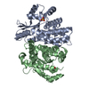
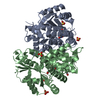
- PDB-3rdp: Crystal structure of thymidine kinase from herpes simplex virus t... -
+ Open data

- Basic information
| Entry | Database: PDB / ID: 3rdp | ||||||
|---|---|---|---|---|---|---|---|
| Title | Crystal structure of thymidine kinase from herpes simplex virus type 1 in complex with N-METHYL-FHBT | ||||||
 Components Components | Thymidine kinase | ||||||
 Keywords Keywords | TRANSFERASE / THYMIDINE KINASE / DNA-SYNTHESIS / PET TRACER / ATP-BINDING / DNA SYNTHESIS / EARLY PROTEIN / NUCLEOTIDE-BINDING | ||||||
| Function / homology |  Function and homology information Function and homology informationTMP biosynthetic process / thymidine kinase / thymidine kinase activity / DNA biosynthetic process / ATP binding Similarity search - Function | ||||||
| Biological species |   Human herpesvirus 1 (Herpes simplex virus type 1) Human herpesvirus 1 (Herpes simplex virus type 1) | ||||||
| Method |  X-RAY DIFFRACTION / SYNCHROTRON / MOLECULAR REPLACEMENT / Resolution: 2.8 Å X-RAY DIFFRACTION / SYNCHROTRON / MOLECULAR REPLACEMENT / Resolution: 2.8 Å | ||||||
 Authors Authors | Pernot, L. / Perozzo, R. / Westermaier, Y. / Martic, M. / Ametamey, S. / Scapozza, L. | ||||||
 Citation Citation | Journal: Nucleosides Nucleotides Nucleic Acids / Year: 2011 Title: Synthesis, crystal structure, and in vitro biological evaluation of C-6 pyrimidine derivatives: new lead structures for monitoring gene expression in vivo. Authors: Martic, M. / Pernot, L. / Westermaier, Y. / Perozzo, R. / Kraljevic, T.G. / Kristafor, S. / Raic-Malic, S. / Scapozza, L. / Ametamey, S. #1: Journal: Proteins / Year: 2000Title: Nucleoside binding site of herpes simplex type 1 thymidine kinase analyzed by X-ray crystallography. Authors: Vogt, J. / Perozzo, R. / Pautsch, A. / Prota, A. / Schelling, P. / Pilger, B. / Folkers, G. / Scapozza, L. / Schulz, G.E. | ||||||
| History |
|
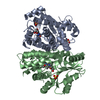
- Structure visualization
| Structure viewer | Molecule: MolmilJmol/JSmol |
|---|
- Downloads & links
-Download
| PDBx/mmCIF format | 3rdp.cif.gz | 134 KB | Display | PDBx/mmCIF format |
|---|---|---|---|---|
| PDB format | pdb3rdp.ent.gz | 104.8 KB | Display | PDB format |
| PDBx/mmJSON format | 3rdp.json.gz | Tree view | PDBx/mmJSON format | |
| Others |  Other downloads Other downloads |
-Validation report
| Summary document | 3rdp_validation.pdf.gz | 468.7 KB | Display | wwPDB validaton report |
|---|---|---|---|---|
| Full document | 3rdp_full_validation.pdf.gz | 477.3 KB | Display | |
| Data in XML | 3rdp_validation.xml.gz | 25.4 KB | Display | |
| Data in CIF | 3rdp_validation.cif.gz | 34.1 KB | Display | |
| Arichive directory | https://data.pdbj.org/pub/pdb/validation_reports/rd/3rdpftp://data.pdbj.org/pub/pdb/validation_reports/rd/3rdp | HTTPS FTP |
-Related structure data
| Related structure data |  3f0tSC S: Starting model for refinement C: citing same article ( |
|---|---|
| Similar structure data |


-Links
 PDBj
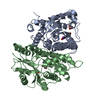
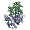
PDBj- Assembly
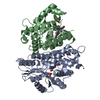
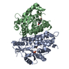
| Deposited unit | 
| ||||||||
|---|---|---|---|---|---|---|---|---|---|
| 1 |
| ||||||||
| Unit cell |
|
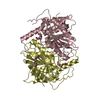
-Components
| #1: Protein | Mass: 35779.086 Da / Num. of mol.: 2 / Fragment: UNP RESIDUES 46-376 Source method: isolated from a genetically manipulated source Source: (gene. exp.) Human herpesvirus 1 (Herpes simplex virus type 1)Strain: strain 17 / Gene: TK, UL23 / Plasmid: pGEX-6P2 / Production host:  References: UniProt: P03176, UniProt: P0DTH5*PLUS, thymidine kinase #2: Chemical |   Mass: 96.063 Da / Num. of mol.: 3 / Source method: obtained synthetically / Formula: SO4 Mass: 96.063 Da / Num. of mol.: 3 / Source method: obtained synthetically / Formula: SO4#3: Chemical | 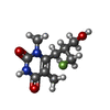  Mass: 230.236 Da / Num. of mol.: 2 / Source method: obtained synthetically / Formula: C10H15FN2O3 Mass: 230.236 Da / Num. of mol.: 2 / Source method: obtained synthetically / Formula: C10H15FN2O3#4: Water | ChemComp-HOH / |  Mass: 18.015 Da / Num. of mol.: 81 / Source method: isolated from a natural source / Formula: H2O Mass: 18.015 Da / Num. of mol.: 81 / Source method: isolated from a natural source / Formula: H2O |
|---|
-Experimental details
-Experiment
| Experiment | Method: X-RAY DIFFRACTION / Number of used crystals: 1 |
|---|
- Sample preparation
| Crystal | Density Matthews: 2.57 Å3/Da / Density % sol: 52.11 % |
|---|---|
| Crystal grow | Temperature: 296 K / Method: vapor diffusion, sitting drop / pH: 7.5 Details: 0.9-1.2M LI2SO4, 1MM DTT, 0.1M HEPES PH 7.5-8.0, VAPOR DIFFUSION, SITTING DROP, temperature 296K |
-Data collection
| Diffraction | Mean temperature: 100 K |
|---|---|
| Diffraction source | Source: SYNCHROTRON / Site: SLS  / Beamline: X06DA / Wavelength: 0.9998 Å / Beamline: X06DA / Wavelength: 0.9998 Å |
| Detector | Type: MARMOSAIC 225 mm CCD / Detector: CCD / Date: Jul 17, 2008 |
| Radiation | Protocol: SINGLE WAVELENGTH / Monochromatic (M) / Laue (L): M / Scattering type: x-ray |
| Radiation wavelength | Wavelength: 0.9998 Å / Relative weight: 1 |
| Reflection | Resolution: 2.8→35.267 Å / Num. obs: 16788 / % possible obs: 91.3 % / Observed criterion σ(I): 2 / Redundancy: 3.6 % / Biso Wilson estimate: 46.19 Å2 / Rmerge(I) obs: 0.218 / Rsym value: 0.186 / Net I/σ(I): 5.94 |
| Reflection shell | Resolution: 2.8→2.95 Å / Redundancy: 3.6 % / Rmerge(I) obs: 0.636 / Mean I/σ(I) obs: 2.3 / Num. unique all: 2475 / Rsym value: 0.566 / % possible all: 93 |
- Processing
| Software |
| ||||||||||||||||||||||||||||||||||||||||||
|---|---|---|---|---|---|---|---|---|---|---|---|---|---|---|---|---|---|---|---|---|---|---|---|---|---|---|---|---|---|---|---|---|---|---|---|---|---|---|---|---|---|---|---|
| Refinement | Method to determine structure: MOLECULAR REPLACEMENT Starting model: 3F0T Resolution: 2.8→35.26 Å / SU ML: 0.37 / Cross valid method: THROUGHOUT / σ(F): 1.38 / Stereochemistry target values: ML
| ||||||||||||||||||||||||||||||||||||||||||
| Solvent computation | Shrinkage radii: 0.83 Å / VDW probe radii: 1.1 Å / Solvent model: FLAT BULK SOLVENT MODEL / Bsol: 18.94 Å2 / ksol: 0.332 e/Å3 | ||||||||||||||||||||||||||||||||||||||||||
| Displacement parameters | Biso mean: 36.35 Å2
| ||||||||||||||||||||||||||||||||||||||||||
| Refinement step | Cycle: LAST / Resolution: 2.8→35.26 Å
| ||||||||||||||||||||||||||||||||||||||||||
| Refine LS restraints |
| ||||||||||||||||||||||||||||||||||||||||||
| LS refinement shell | Refine-ID: X-RAY DIFFRACTION / Total num. of bins used: 6
|
