 Movie
Movie Controller

[English] 日本語
 Yorodumi
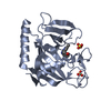
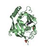
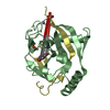
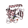
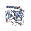
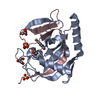
- PDB-3p0p: Human Tankyrase 2 - Catalytic PARP domain in complex with an inhibitor -
+ Open data

- Basic information
| Entry | Database: PDB / ID: 3p0p | ||||||
|---|---|---|---|---|---|---|---|
| Title | Human Tankyrase 2 - Catalytic PARP domain in complex with an inhibitor | ||||||
 Components Components | Tankyrase-2 | ||||||
 Keywords Keywords | TRANSFERASE/TRANSFERASE INHIBITOR / PROTEIN-LIGAND COMPLEX / Structural Genomics / Structural Genomics Consortium / SGC / DIPHTHERIA TOXIN LIKE FOLD / TRANSFERASE / NAD+ / ADP-RIBOSYLATION / TRANSFERASE-TRANSFERASE INHIBITOR complex | ||||||
| Function / homology |  Function and homology information Function and homology informationXAV939 stabilizes AXIN / positive regulation of telomere capping / NAD+ ADP-ribosyltransferase / protein auto-ADP-ribosylation / protein localization to chromosome, telomeric region / negative regulation of telomere maintenance via telomere lengthening / NAD+-protein-aspartate ADP-ribosyltransferase activity / protein poly-ADP-ribosylation / NAD+-protein-glutamate ADP-ribosyltransferase activity / NAD+-protein mono-ADP-ribosyltransferase activity ...XAV939 stabilizes AXIN / positive regulation of telomere capping / NAD+ ADP-ribosyltransferase / protein auto-ADP-ribosylation / protein localization to chromosome, telomeric region / negative regulation of telomere maintenance via telomere lengthening / NAD+-protein-aspartate ADP-ribosyltransferase activity / protein poly-ADP-ribosylation / NAD+-protein-glutamate ADP-ribosyltransferase activity / NAD+-protein mono-ADP-ribosyltransferase activity / pericentriolar material / Transferases; Glycosyltransferases; Pentosyltransferases / NAD+ poly-ADP-ribosyltransferase activity / positive regulation of telomere maintenance via telomerase / nucleotidyltransferase activity / TCF dependent signaling in response to WNT / Degradation of AXIN / Regulation of PTEN stability and activity / Wnt signaling pathway / protein polyubiquitination / nuclear envelope / positive regulation of canonical Wnt signaling pathway / chromosome, telomeric region / Ub-specific processing proteases / Golgi membrane / perinuclear region of cytoplasm / enzyme binding / metal ion binding / nucleus / cytosol / cytoplasm Similarity search - Function | ||||||
| Biological species |  Homo sapiens (human) Homo sapiens (human) | ||||||
| Method |  X-RAY DIFFRACTION / MOLECULAR REPLACEMENT / Resolution: 2.49 Å X-RAY DIFFRACTION / MOLECULAR REPLACEMENT / Resolution: 2.49 Å | ||||||
 Authors Authors | Karlberg, T. / Siponen, M.I. / Arrowsmith, C.H. / Berglund, H. / Bountra, C. / Collins, R. / Edwards, A.M. / Flodin, S. / Flores, A. / Graslund, S. ...Karlberg, T. / Siponen, M.I. / Arrowsmith, C.H. / Berglund, H. / Bountra, C. / Collins, R. / Edwards, A.M. / Flodin, S. / Flores, A. / Graslund, S. / Hammarstrom, M. / Johansson, I. / Kotenyova, T. / Kouznetsova, E. / Moche, M. / Nordlund, P. / Nyman, T. / Persson, C. / Schutz, P. / Sehic, A. / Thorsell, A.G. / Tresaugues, L. / Van Den Berg, S. / Wahlberg, E. / Weigelt, J. / Welin, M. / Schuler, H. / Structural Genomics Consortium (SGC) | ||||||
 Citation Citation | Journal: Nat.Biotechnol. / Year: 2012 Title: Family-wide chemical profiling and structural analysis of PARP and tankyrase inhibitors Authors: Wahlberg, E. / Karlberg, T. / Kouznetsova, E. / Markova, N. / Macchiarulo, A. / Thorsell, A.G. / Pol, E. / Frostell, A. / Ekblad, T. / Kull, B. / Robertson, G.M. / Pellicciari, R. / Schuler, H. / Weigelt, J. | ||||||
| History |
|
- Structure visualization
| Structure viewer | Molecule: MolmilJmol/JSmol |
|---|
- Downloads & links
-Download
| PDBx/mmCIF format | 3p0p.cif.gz | 102.3 KB | Display | PDBx/mmCIF format |
|---|---|---|---|---|
| PDB format | pdb3p0p.ent.gz | 76.2 KB | Display | PDB format |
| PDBx/mmJSON format | 3p0p.json.gz | Tree view | PDBx/mmJSON format | |
| Others |  Other downloads Other downloads |
-Validation report
| Arichive directory | https://data.pdbj.org/pub/pdb/validation_reports/p0/3p0pftp://data.pdbj.org/pub/pdb/validation_reports/p0/3p0p | HTTPS FTP |
|---|
-Related structure data
| Related structure data | 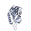 3goyC 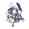 3mhjC 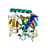 3mhkC 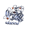 3p0nC 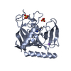 3p0qC  3se2C  3smiC 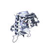 3smjC 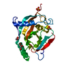 3kr7S S: Starting model for refinement C: citing same article ( |
|---|---|
| Similar structure data |


-Links
 PDBj
PDBj


- Assembly
| Deposited unit | 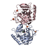
| ||||||||
|---|---|---|---|---|---|---|---|---|---|
| 1 | 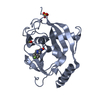
| ||||||||
| 2 | 
| ||||||||
| Unit cell |
|
-Components
| #1: Protein | Mass: 27299.764 Da / Num. of mol.: 2 / Fragment: Catalytic domain Source method: isolated from a genetically manipulated source Source: (gene. exp.) Homo sapiens (human) / Gene: TNKS2 / Plasmid: pNIC-Bsa4 / Production host:  #2: Chemical |   Mass: 65.409 Da / Num. of mol.: 2 / Source method: obtained synthetically / Formula: Zn Mass: 65.409 Da / Num. of mol.: 2 / Source method: obtained synthetically / Formula: Zn#3: Chemical | 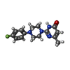  Mass: 288.320 Da / Num. of mol.: 2 / Source method: obtained synthetically / Formula: C15H17FN4O Mass: 288.320 Da / Num. of mol.: 2 / Source method: obtained synthetically / Formula: C15H17FN4O#4: Chemical | ChemComp-SO4 /   Mass: 96.063 Da / Num. of mol.: 4 / Source method: obtained synthetically / Formula: SO4 Mass: 96.063 Da / Num. of mol.: 4 / Source method: obtained synthetically / Formula: SO4#5: Water | ChemComp-HOH / |  Mass: 18.015 Da / Num. of mol.: 115 / Source method: isolated from a natural source / Formula: H2O Mass: 18.015 Da / Num. of mol.: 115 / Source method: isolated from a natural source / Formula: H2O |
|---|
-Experimental details
-Experiment
| Experiment | Method: X-RAY DIFFRACTION / Number of used crystals: 1 |
|---|
- Sample preparation
| Crystal | Density Matthews: 2.4 Å3/Da / Density % sol: 48.78 % |
|---|---|
| Crystal grow | Temperature: 277 K / Method: vapor diffusion, sitting drop / pH: 8 Details: 23% PEG3350, 0.2M ammonium sulfate, 0.1M Tris-HCl, pH 8, VAPOR DIFFUSION, SITTING DROP, temperature 277K |
-Data collection
| Diffraction | Mean temperature: 100 K |
|---|---|
| Diffraction source | Source: ROTATING ANODE / Type: RIGAKU / Wavelength: 1.54177 Å |
| Detector | Type: Bruker Platinum 135 / Detector: CCD / Date: Feb 4, 2010 |
| Radiation | Monochromator: graphite / Protocol: SINGLE WAVELENGTH / Monochromatic (M) / Laue (L): M / Scattering type: x-ray |
| Radiation wavelength | Wavelength: 1.54177 Å / Relative weight: 1 |
| Reflection | Resolution: 2.49→66 Å / Num. all: 18543 / Num. obs: 18543 / % possible obs: 99 % / Observed criterion σ(F): 0 / Observed criterion σ(I): 0 / Redundancy: 6.6 % / Biso Wilson estimate: 34.38 Å2 / Rmerge(I) obs: 0.1855 / Rsym value: 0.1186 / Net I/σ(I): 7.6 |
| Reflection shell | Resolution: 2.49→2.59 Å / Redundancy: 4 % / Rmerge(I) obs: 0.3858 / Mean I/σ(I) obs: 2.5 / Num. unique all: 1813 / Rsym value: 0.4221 / % possible all: 91.5 |
- Processing
| Software |
| ||||||||||||||||||||||||||||||||||||||||||||||||||||||||||||||||||||||||||||||||||||||||||||||||||||||||||||
|---|---|---|---|---|---|---|---|---|---|---|---|---|---|---|---|---|---|---|---|---|---|---|---|---|---|---|---|---|---|---|---|---|---|---|---|---|---|---|---|---|---|---|---|---|---|---|---|---|---|---|---|---|---|---|---|---|---|---|---|---|---|---|---|---|---|---|---|---|---|---|---|---|---|---|---|---|---|---|---|---|---|---|---|---|---|---|---|---|---|---|---|---|---|---|---|---|---|---|---|---|---|---|---|---|---|---|---|---|---|
| Refinement | Method to determine structure: MOLECULAR REPLACEMENT Starting model: PDB ENTRY 3KR7 Resolution: 2.49→24.86 Å / Cor.coef. Fo:Fc: 0.9063 / Cor.coef. Fo:Fc free: 0.8217 / Occupancy max: 1 / Occupancy min: 0.5 / Cross valid method: THROUGHOUT / σ(F): 0 / σ(I): 0 / Stereochemistry target values: MAXIMUM LIKELIHOOD
| ||||||||||||||||||||||||||||||||||||||||||||||||||||||||||||||||||||||||||||||||||||||||||||||||||||||||||||
| Displacement parameters | Biso max: 101.76 Å2 / Biso mean: 25.38 Å2 / Biso min: 2.44 Å2
| ||||||||||||||||||||||||||||||||||||||||||||||||||||||||||||||||||||||||||||||||||||||||||||||||||||||||||||
| Refine analyze | Luzzati coordinate error obs: 0.304 Å | ||||||||||||||||||||||||||||||||||||||||||||||||||||||||||||||||||||||||||||||||||||||||||||||||||||||||||||
| Refinement step | Cycle: LAST / Resolution: 2.49→24.86 Å
| ||||||||||||||||||||||||||||||||||||||||||||||||||||||||||||||||||||||||||||||||||||||||||||||||||||||||||||
| Refine LS restraints |
| ||||||||||||||||||||||||||||||||||||||||||||||||||||||||||||||||||||||||||||||||||||||||||||||||||||||||||||
| LS refinement shell | Resolution: 2.49→2.64 Å / Total num. of bins used: 9
|
