 Movie
Movie Controller


+ Open data

- Basic information
| Entry | Database: PDB / ID: 3lzt | ||||||
|---|---|---|---|---|---|---|---|
| Title | REFINEMENT OF TRICLINIC LYSOZYME AT ATOMIC RESOLUTION | ||||||
 Components Components | LYSOZYME | ||||||
 Keywords Keywords | HYDROLASE / O-GLYCOSYL / GLYCOSIDASE | ||||||
| Function / homology |  Function and homology information Function and homology informationLactose synthesis / Antimicrobial peptides / Neutrophil degranulation / beta-N-acetylglucosaminidase activity / cell wall macromolecule catabolic process / lysozyme / lysozyme activity / killing of cells of another organism / defense response to Gram-negative bacterium / defense response to bacterium ...Lactose synthesis / Antimicrobial peptides / Neutrophil degranulation / beta-N-acetylglucosaminidase activity / cell wall macromolecule catabolic process / lysozyme / lysozyme activity / killing of cells of another organism / defense response to Gram-negative bacterium / defense response to bacterium / defense response to Gram-positive bacterium / endoplasmic reticulum / : / identical protein binding / cytoplasm Similarity search - Function | ||||||
| Biological species |  | ||||||
| Method |  X-RAY DIFFRACTION / SYNCHROTRON / MOLECULAR REPLACEMENT / Resolution: 0.925 Å X-RAY DIFFRACTION / SYNCHROTRON / MOLECULAR REPLACEMENT / Resolution: 0.925 Å | ||||||
 Authors Authors | Walsh, M.A. / Schneider, T. / Sieker, L.C. / Dauter, Z. / Lamzin, V. / Wilson, K.S. | ||||||
 Citation Citation | Journal: Acta Crystallogr.,Sect.D / Year: 1998 Title: Refinement of triclinic hen egg-white lysozyme at atomic resolution. Authors: Walsh, M.A. / Schneider, T.R. / Sieker, L.C. / Dauter, Z. / Lamzin, V.S. / Wilson, K.S. #1: Journal: Acta Crystallogr.,Sect.B / Year: 1990Title: Refinement of Triclinic Lysozyme: I. Fourier and Least-Squares Methods Authors: Hodsdon, J.M. / Brown, G.M. / Sieker, L.C. / Jensen, L.H. #2: Journal: Acta Crystallogr.,Sect.B / Year: 1990Title: Refinement of Triclinic Lysozyme: II. The Method of Stereochemically Restrained Least Squares Authors: Ramanadham, M. / Sieker, L.C. / Jensen, L.H. | ||||||
| History |
|
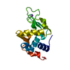
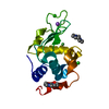
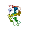
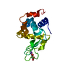
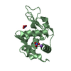
- Structure visualization
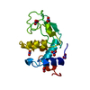
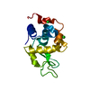
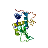
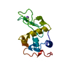
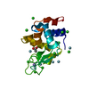
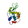
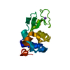
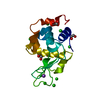
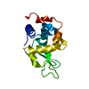
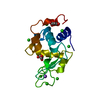
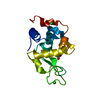
| Structure viewer | Molecule: MolmilJmol/JSmol |
|---|
- Downloads & links
-Download
| PDBx/mmCIF format | 3lzt.cif.gz | 81.9 KB | Display | PDBx/mmCIF format |
|---|---|---|---|---|
| PDB format | pdb3lzt.ent.gz | 60.8 KB | Display | PDB format |
| PDBx/mmJSON format | 3lzt.json.gz | Tree view | PDBx/mmJSON format | |
| Others |  Other downloads Other downloads |
-Validation report
| Arichive directory | https://data.pdbj.org/pub/pdb/validation_reports/lz/3lztftp://data.pdbj.org/pub/pdb/validation_reports/lz/3lzt | HTTPS FTP |
|---|
-Related structure data


-Links
 PDBj
PDBj


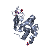
- Assembly
| Deposited unit | 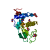
| ||||||||
|---|---|---|---|---|---|---|---|---|---|
| 1 |
| ||||||||
| Unit cell |
|
-Components
| #1: Protein | Mass: 14331.160 Da / Num. of mol.: 1 / Source method: isolated from a natural source / Details: NITRATE AND ACETATE IONS PRESENT / Source: (natural) | ||||||
|---|---|---|---|---|---|---|---|
| #2: Chemical | ChemComp-NO3 /   Mass: 62.005 Da / Num. of mol.: 6 / Source method: obtained synthetically / Formula: NO3 Mass: 62.005 Da / Num. of mol.: 6 / Source method: obtained synthetically / Formula: NO3#3: Chemical |   Mass: 59.044 Da / Num. of mol.: 3 / Source method: obtained synthetically / Formula: C2H3O2 Mass: 59.044 Da / Num. of mol.: 3 / Source method: obtained synthetically / Formula: C2H3O2#4: Water | ChemComp-HOH / |  Mass: 18.015 Da / Num. of mol.: 245 / Source method: isolated from a natural source / Formula: H2O Mass: 18.015 Da / Num. of mol.: 245 / Source method: isolated from a natural source / Formula: H2OHas protein modification | Y | |
-Experimental details
-Experiment
| Experiment | Method: X-RAY DIFFRACTION / Number of used crystals: 1 |
|---|
- Sample preparation
| Crystal | Density Matthews: 1.69 Å3/Da / Density % sol: 26.9 % | ||||||||||||||||||||
|---|---|---|---|---|---|---|---|---|---|---|---|---|---|---|---|---|---|---|---|---|---|
| Crystal grow | Method: batch method / pH: 4.6 Details: BATCH METHOD USED. 1% PROTEIN SOLUTION IN 100MM SODIUM ACETATE PH 4.5-4.6. SODIUM NITRATE ADDED TO A CONCENTRATION OF 20MGS/ML. CRYSTALS GROWN AT ROOM TEMPERATURE., batch method PH range: 4.5-4.6 / Temp details: room temp | ||||||||||||||||||||
| Crystal grow | *PLUS Temperature: 296 K / pH: 4.5 / Method: unknown | ||||||||||||||||||||
| Components of the solutions | *PLUS
|
-Data collection
| Diffraction | Mean temperature: 120 K |
|---|---|
| Diffraction source | Source: SYNCHROTRON / Site: EMBL/DESY, HAMBURG  / Beamline: X11 / Wavelength: 0.927 / Beamline: X11 / Wavelength: 0.927 |
| Detector | Type: MARRESEARCH / Detector: IMAGE PLATE / Date: Dec 1, 1994 / Details: SEGMENTED MIRROR |
| Radiation | Monochromator: SI(111) / Monochromatic (M) / Laue (L): M / Scattering type: x-ray |
| Radiation wavelength | Wavelength: 0.927 Å / Relative weight: 1 |
| Reflection | Resolution: 0.925→25 Å / Num. obs: 58373 / % possible obs: 90.1 % / Observed criterion σ(I): 3 / Redundancy: 2 % / Rmerge(I) obs: 0.028 / Rsym value: 0.028 / Net I/σ(I): 29.1 |
| Reflection shell | Resolution: 0.925→0.94 Å / Redundancy: 2 % / Rmerge(I) obs: 0.17 / Mean I/σ(I) obs: 4.9 / Rsym value: 0.17 / % possible all: 78 |
| Reflection | *PLUS Num. measured all: 232156 |
| Reflection shell | *PLUS % possible obs: 78 % |
- Processing
| Software |
| |||||||||||||||||||||||||||||||||
|---|---|---|---|---|---|---|---|---|---|---|---|---|---|---|---|---|---|---|---|---|---|---|---|---|---|---|---|---|---|---|---|---|---|---|
| Refinement | Method to determine structure: MOLECULAR REPLACEMENT / Resolution: 0.925→20 Å / Num. parameters: 12844 / Num. restraintsaints: 16207 / Cross valid method: FREE R / σ(F): 0 / Stereochemistry target values: ENGH AND HUBER Details: WATER OCCUPANCIES REFINED. TWO SECTIONS OF THE MAIN CHAIN MODELED IN TWO DISCRETE CONFORMATIONS. DISORDERED REGIONS WERE MODELED STEREOCHEMICALLY.
| |||||||||||||||||||||||||||||||||
| Solvent computation | Solvent model: MOEWS & KRETSINGER | |||||||||||||||||||||||||||||||||
| Refine analyze | Num. disordered residues: 28 / Occupancy sum hydrogen: 923.71 / Occupancy sum non hydrogen: 1230.1 | |||||||||||||||||||||||||||||||||
| Refinement step | Cycle: LAST / Resolution: 0.925→20 Å
| |||||||||||||||||||||||||||||||||
| Refine LS restraints |
| |||||||||||||||||||||||||||||||||
| Software | *PLUS Name: SHELXL-96 / Classification: refinement | |||||||||||||||||||||||||||||||||
| Refinement | *PLUS Rfactor obs: 0.093 | |||||||||||||||||||||||||||||||||
| Solvent computation | *PLUS | |||||||||||||||||||||||||||||||||
| Displacement parameters | *PLUS | |||||||||||||||||||||||||||||||||
| Refine LS restraints | *PLUS
|
