 Movie
Movie Controller

[English] 日本語
 Yorodumi
- PDB-3lwl: Structure of Klenow fragment of Taq polymerase in complex with an... -
+ Open data

- Basic information
| Entry | Database: PDB / ID: 3lwl | ||||||
|---|---|---|---|---|---|---|---|
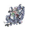
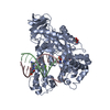
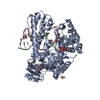
| Title | Structure of Klenow fragment of Taq polymerase in complex with an abasic site | ||||||
 Components Components |
| ||||||
 Keywords Keywords | TRANSFERASE/DNA / DNA replication / DNA repair / DNA polymerases / Abasic sites / Translesion synthesis / DNA damage / DNA-binding / DNA-directed DNA polymerase / amino acid-templating mechanism / TRANSFERASE-DNA complex | ||||||
| Function / homology |  Function and homology information Function and homology informationnucleoside binding / 5'-3' exonuclease activity / DNA-templated DNA replication / double-strand break repair / DNA-directed DNA polymerase / DNA-directed DNA polymerase activity / DNA binding Similarity search - Function | ||||||
| Biological species |   Thermus aquaticus (bacteria) Thermus aquaticus (bacteria) | ||||||
| Method |  X-RAY DIFFRACTION / SYNCHROTRON / MOLECULAR REPLACEMENT / Resolution: 2.25 Å X-RAY DIFFRACTION / SYNCHROTRON / MOLECULAR REPLACEMENT / Resolution: 2.25 Å | ||||||
 Authors Authors | Marx, A. / Diederichs, K. / Obeid, S. | ||||||
 Citation Citation | Journal: Embo J. / Year: 2010 Title: Replication through an abasic DNA lesion: structural basis for adenine selectivity Authors: Obeid, S. / Blatter, N. / Kranaster, R. / Schnur, A. / Diederichs, K. / Welte, W. / Marx, A. | ||||||
| History |
|
- Structure visualization
| Structure viewer | Molecule: MolmilJmol/JSmol |
|---|
- Downloads & links
-Download
| PDBx/mmCIF format | 3lwl.cif.gz | 267.6 KB | Display | PDBx/mmCIF format |
|---|---|---|---|---|
| PDB format | pdb3lwl.ent.gz | 211.4 KB | Display | PDB format |
| PDBx/mmJSON format | 3lwl.json.gz | Tree view | PDBx/mmJSON format | |
| Others |  Other downloads Other downloads |
-Validation report
| Arichive directory | https://data.pdbj.org/pub/pdb/validation_reports/lw/3lwlftp://data.pdbj.org/pub/pdb/validation_reports/lw/3lwl | HTTPS FTP |
|---|
-Related structure data
| Related structure data |  3lwmC  3ktqS S: Starting model for refinement C: citing same article ( |
|---|---|
| Similar structure data |


-Links
 PDBj
PDBj


- Assembly
| Deposited unit | 
| ||||||||||||
|---|---|---|---|---|---|---|---|---|---|---|---|---|---|
| 1 |
| ||||||||||||
| Unit cell |
| ||||||||||||
| Components on special symmetry positions |
|
-Components
-Protein , 1 types, 1 molecules A
| #1: Protein | Mass: 60936.965 Da / Num. of mol.: 1 / Fragment: KLENOW FRAGMENT, UNP residues 293-832 Source method: isolated from a genetically manipulated source Source: (gene. exp.) Thermus aquaticus (bacteria) / Gene: pol1, polA / Plasmid: pET-21b / Production host: |
|---|
-DNA chain , 2 types, 2 molecules BC
| #2: DNA chain | Mass: 3641.395 Da / Num. of mol.: 1 / Source method: obtained synthetically / Details: Synthetic DNA |
|---|---|
| #3: DNA chain | Mass: 4790.093 Da / Num. of mol.: 1 / Source method: obtained synthetically / Details: Synthetic DNA |
-Non-polymers , 6 types, 179 molecules 


| #4: Chemical | ChemComp-DDS /  Mass: 475.182 Da / Num. of mol.: 1 / Source method: obtained synthetically / Formula: C10H16N5O11P3 Mass: 475.182 Da / Num. of mol.: 1 / Source method: obtained synthetically / Formula: C10H16N5O11P3 | ||||||
|---|---|---|---|---|---|---|---|
| #5: Chemical | ChemComp-MG /  Mass: 24.305 Da / Num. of mol.: 1 / Source method: obtained synthetically / Formula: Mg Mass: 24.305 Da / Num. of mol.: 1 / Source method: obtained synthetically / Formula: Mg | ||||||
| #6: Chemical | ChemComp-ACT /  Mass: 59.044 Da / Num. of mol.: 5 / Source method: obtained synthetically / Formula: C2H3O2 Mass: 59.044 Da / Num. of mol.: 5 / Source method: obtained synthetically / Formula: C2H3O2#7: Chemical | ChemComp-GOL /  Mass: 92.094 Da / Num. of mol.: 7 / Source method: obtained synthetically / Formula: C3H8O3 Mass: 92.094 Da / Num. of mol.: 7 / Source method: obtained synthetically / Formula: C3H8O3#8: Chemical | ChemComp-NA /  Mass: 22.990 Da / Num. of mol.: 4 / Source method: obtained synthetically / Formula: Na Mass: 22.990 Da / Num. of mol.: 4 / Source method: obtained synthetically / Formula: Na#9: Water | ChemComp-HOH / | Mass: 18.015 Da / Num. of mol.: 161 / Source method: isolated from a natural source / Formula: H2O |
-Experimental details
-Experiment
| Experiment | Method: X-RAY DIFFRACTION / Number of used crystals: 1 |
|---|
- Sample preparation
| Crystal | Density Matthews: 2.31 Å3/Da / Density % sol: 46.69 % |
|---|---|
| Crystal grow | Temperature: 291 K / Method: vapor diffusion, hanging drop / pH: 7 Details: 0.05M sodium cacodylate, 0.2M ammonium acetate, 0.01M magnesium acetate, 30% PEG 8000, pH 7, VAPOR DIFFUSION, HANGING DROP, temperature 291K |
-Data collection
| Diffraction | Mean temperature: 100 K |
|---|---|
| Diffraction source | Source: SYNCHROTRON / Site: SLS  / Beamline: X06DA / Wavelength: 1 Å / Beamline: X06DA / Wavelength: 1 Å |
| Detector | Type: MARMOSAIC 225 mm CCD / Detector: CCD / Date: Sep 18, 2009 Details: vertically collimating mirror (M1, focus at infinity), followed by a Bartels Monochromator with dual channel cut crystals |
| Radiation | Monochromator: Bartels Monochromator with dual channel cut crystals (DCCM) in (+--+) geometry, and a toroidal mirror (M2) Protocol: SINGLE WAVELENGTH / Monochromatic (M) / Laue (L): M / Scattering type: x-ray |
| Radiation wavelength | Wavelength: 1 Å / Relative weight: 1 |
| Reflection | Resolution: 2.25→50 Å / Num. obs: 30775 / % possible obs: 99.7 % / Observed criterion σ(F): 0 / Observed criterion σ(I): -3 / Redundancy: 10.78 % / Biso Wilson estimate: 43.283 Å2 / Rmerge(I) obs: 0.124 / Net I/σ(I): 16.11 |
| Reflection shell | Resolution: 2.25→2.38 Å / Redundancy: 9.94 % / Rmerge(I) obs: 0.971 / Mean I/σ(I) obs: 2.48 / Num. unique all: 4843 / % possible all: 98.5 |
- Processing
| Software |
| ||||||||||||||||||||||||||||||||||||||||||||||||||||||||||||||||||||||||||||||||||||||||||||||||||||||||||||||||||||||||||||||||||||||||||||||||||||||
|---|---|---|---|---|---|---|---|---|---|---|---|---|---|---|---|---|---|---|---|---|---|---|---|---|---|---|---|---|---|---|---|---|---|---|---|---|---|---|---|---|---|---|---|---|---|---|---|---|---|---|---|---|---|---|---|---|---|---|---|---|---|---|---|---|---|---|---|---|---|---|---|---|---|---|---|---|---|---|---|---|---|---|---|---|---|---|---|---|---|---|---|---|---|---|---|---|---|---|---|---|---|---|---|---|---|---|---|---|---|---|---|---|---|---|---|---|---|---|---|---|---|---|---|---|---|---|---|---|---|---|---|---|---|---|---|---|---|---|---|---|---|---|---|---|---|---|---|---|---|---|---|
| Refinement | Method to determine structure: MOLECULAR REPLACEMENT Starting model: PDB ENTRY 3KTQ Resolution: 2.25→47.73 Å / Occupancy max: 1 / Occupancy min: 0 / FOM work R set: 0.836 / SU ML: 0.28 / Cross valid method: THROUGHOUT / σ(F): 0 / σ(I): -3 / Phase error: 23.46 / Stereochemistry target values: ML
| ||||||||||||||||||||||||||||||||||||||||||||||||||||||||||||||||||||||||||||||||||||||||||||||||||||||||||||||||||||||||||||||||||||||||||||||||||||||
| Solvent computation | Shrinkage radii: 0.9 Å / VDW probe radii: 1.11 Å / Solvent model: FLAT BULK SOLVENT MODEL / Bsol: 42.922 Å2 / ksol: 0.345 e/Å3 | ||||||||||||||||||||||||||||||||||||||||||||||||||||||||||||||||||||||||||||||||||||||||||||||||||||||||||||||||||||||||||||||||||||||||||||||||||||||
| Displacement parameters | Biso max: 291.28 Å2 / Biso mean: 52.624 Å2 / Biso min: 15.86 Å2
| ||||||||||||||||||||||||||||||||||||||||||||||||||||||||||||||||||||||||||||||||||||||||||||||||||||||||||||||||||||||||||||||||||||||||||||||||||||||
| Refine analyze | Luzzati coordinate error obs: 0.28 Å | ||||||||||||||||||||||||||||||||||||||||||||||||||||||||||||||||||||||||||||||||||||||||||||||||||||||||||||||||||||||||||||||||||||||||||||||||||||||
| Refinement step | Cycle: LAST / Resolution: 2.25→47.73 Å
| ||||||||||||||||||||||||||||||||||||||||||||||||||||||||||||||||||||||||||||||||||||||||||||||||||||||||||||||||||||||||||||||||||||||||||||||||||||||
| Refine LS restraints |
| ||||||||||||||||||||||||||||||||||||||||||||||||||||||||||||||||||||||||||||||||||||||||||||||||||||||||||||||||||||||||||||||||||||||||||||||||||||||
| LS refinement shell |
| ||||||||||||||||||||||||||||||||||||||||||||||||||||||||||||||||||||||||||||||||||||||||||||||||||||||||||||||||||||||||||||||||||||||||||||||||||||||
| Refinement TLS params. | Method: refined / Refine-ID: X-RAY DIFFRACTION
| ||||||||||||||||||||||||||||||||||||||||||||||||||||||||||||||||||||||||||||||||||||||||||||||||||||||||||||||||||||||||||||||||||||||||||||||||||||||
| Refinement TLS group |
|
