 Movie
Movie Controller

[English] 日本語
 Yorodumi
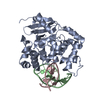
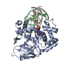
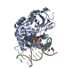
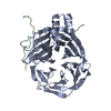
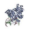
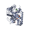
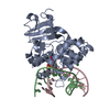
- PDB-3ktu: Structure of human 8-oxoGuanine Glycosylase 1 bound to fluorninat... -
+ Open data

- Basic information
| Entry | Database: PDB / ID: 3ktu | ||||||
|---|---|---|---|---|---|---|---|
| Title | Structure of human 8-oxoGuanine Glycosylase 1 bound to fluorninated oxoG-containing DNA | ||||||
 Components Components |
| ||||||
 Keywords Keywords | HYDROLASE / LYASE/DNA / 8-oxoGuanine / 2'-fluoro-8-oxoGuanine / protein-DNA complex / DNA glycosylase / Base Excsion Repair / DNA damage / DNA repair / LYASE-DNA complex | ||||||
| Function / homology |  Function and homology information Function and homology informationDefective OGG1 Substrate Binding / Defective OGG1 Substrate Processing / Defective OGG1 Localization / negative regulation of double-strand break repair via single-strand annealing / depurination / oxidized purine nucleobase lesion DNA N-glycosylase activity / base-excision repair, AP site formation / depyrimidination / 8-oxo-7,8-dihydroguanine DNA N-glycosylase activity / Displacement of DNA glycosylase by APEX1 ...Defective OGG1 Substrate Binding / Defective OGG1 Substrate Processing / Defective OGG1 Localization / negative regulation of double-strand break repair via single-strand annealing / depurination / oxidized purine nucleobase lesion DNA N-glycosylase activity / base-excision repair, AP site formation / depyrimidination / 8-oxo-7,8-dihydroguanine DNA N-glycosylase activity / Displacement of DNA glycosylase by APEX1 / positive regulation of gene expression via chromosomal CpG island demethylation / response to folic acid / oxidized purine DNA binding / Hydrolases; Glycosylases; Hydrolysing N-glycosyl compounds / APEX1-Independent Resolution of AP Sites via the Single Nucleotide Replacement Pathway / response to light stimulus / Recognition and association of DNA glycosylase with site containing an affected purine / Cleavage of the damaged purine / Recognition and association of DNA glycosylase with site containing an affected pyrimidine / Cleavage of the damaged pyrimidine / cellular response to cadmium ion / class I DNA-(apurinic or apyrimidinic site) endonuclease activity / DNA-(apurinic or apyrimidinic site) lyase / nucleotide-excision repair / base-excision repair / response to radiation / nuclear matrix / cellular response to reactive oxygen species / response to estradiol / microtubule binding / endonuclease activity / response to ethanol / response to oxidative stress / damaged DNA binding / nuclear speck / mitochondrial matrix / response to xenobiotic stimulus / RNA polymerase II cis-regulatory region sequence-specific DNA binding / DNA damage response / regulation of DNA-templated transcription / negative regulation of apoptotic process / enzyme binding / positive regulation of transcription by RNA polymerase II / protein-containing complex / nucleoplasm / nucleus / cytosol Similarity search - Function | ||||||
| Biological species |  Homo sapiens (human) Homo sapiens (human)synthetic construct (others) | ||||||
| Method |  X-RAY DIFFRACTION / SYNCHROTRON / MOLECULAR REPLACEMENT / Resolution: 2.3 Å X-RAY DIFFRACTION / SYNCHROTRON / MOLECULAR REPLACEMENT / Resolution: 2.3 Å | ||||||
 Authors Authors | Verdine, G.L. / Lee, S.M. | ||||||
 Citation Citation | Journal: to be published Title: Structural investigation of hOGG1 bound to a fluorinated oxoG analog Authors: Verdine, G.L. / Lee, S.M. #1: Journal: to be publishedTitle: A Catalytic Checkpoint in Base Excision by the Human 8-Oxoguanine DNA Glycosylase hOGG1 Authors: Verdine, G.L. / Crenshaw, C.M. / Oo, K.S. / Kutchukian, P.S. | ||||||
| History |
|
- Structure visualization
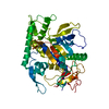
| Structure viewer | Molecule: MolmilJmol/JSmol |
|---|
- Downloads & links
-Download
| PDBx/mmCIF format | 3ktu.cif.gz | 94.1 KB | Display | PDBx/mmCIF format |
|---|---|---|---|---|
| PDB format | pdb3ktu.ent.gz | 67.1 KB | Display | PDB format |
| PDBx/mmJSON format | 3ktu.json.gz | Tree view | PDBx/mmJSON format | |
| Others |  Other downloads Other downloads |
-Validation report
| Summary document | 3ktu_validation.pdf.gz | 440.6 KB | Display | wwPDB validaton report |
|---|---|---|---|---|
| Full document | 3ktu_full_validation.pdf.gz | 448.7 KB | Display | |
| Data in XML | 3ktu_validation.xml.gz | 15.6 KB | Display | |
| Data in CIF | 3ktu_validation.cif.gz | 21.1 KB | Display | |
| Arichive directory | https://data.pdbj.org/pub/pdb/validation_reports/kt/3ktuftp://data.pdbj.org/pub/pdb/validation_reports/kt/3ktu | HTTPS FTP |
-Related structure data
| Related structure data |  1ebmS S: Starting model for refinement |
|---|---|
| Similar structure data |

-Links
 PDBj
PDBj


- Assembly
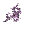
| Deposited unit | 
| ||||||||
|---|---|---|---|---|---|---|---|---|---|
| 1 |
| ||||||||
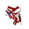
| Unit cell |
|
-Components
| #1: Protein | Mass: 35727.469 Da / Num. of mol.: 1 / Fragment: sequence database residues 12-325 Source method: isolated from a genetically manipulated source Source: (gene. exp.) Homo sapiens (human) / Gene: MMH, MUTM, OGG1, OGH1 / Plasmid: pET30a / Production host:  References: UniProt: O15527, Hydrolases; Glycosylases; Hydrolysing N-glycosyl compounds, DNA-(apurinic or apyrimidinic site) lyase | ||
|---|---|---|---|
| #2: DNA chain | Mass: 4016.623 Da / Num. of mol.: 1 / Source method: obtained synthetically / Details: SYNTHETIC DNA / Source: (synth.) synthetic construct (others) | ||
| #3: DNA chain | Mass: 3672.369 Da / Num. of mol.: 1 / Source method: obtained synthetically / Details: SYNTHETIC DNA / Source: (synth.) synthetic construct (others) | ||
| #4: Chemical |   Mass: 40.078 Da / Num. of mol.: 2 / Source method: obtained synthetically / Formula: Ca Mass: 40.078 Da / Num. of mol.: 2 / Source method: obtained synthetically / Formula: Ca#5: Water | ChemComp-HOH / |  Mass: 18.015 Da / Num. of mol.: 72 / Source method: isolated from a natural source / Formula: H2O Mass: 18.015 Da / Num. of mol.: 72 / Source method: isolated from a natural source / Formula: H2O |
-Experimental details
-Experiment
| Experiment | Method: X-RAY DIFFRACTION / Number of used crystals: 1 |
|---|
- Sample preparation
| Crystal | Density Matthews: 3.03 Å3/Da / Density % sol: 59.39 % | ||||||||||||||||||||||||||||
|---|---|---|---|---|---|---|---|---|---|---|---|---|---|---|---|---|---|---|---|---|---|---|---|---|---|---|---|---|---|
| Crystal grow | Temperature: 277 K / pH: 6 Details: Calcium Chloride, Sodium Cacodylate, pH 6.0, PEG 8000, VAPOR DIFFUSION, HANGING DROP, temperature 277K | ||||||||||||||||||||||||||||
| Components of the solutions |
|
-Data collection
| Diffraction | Mean temperature: 100 K |
|---|---|
| Diffraction source | Source: SYNCHROTRON / Site: APS  / Beamline: 24-ID-C / Wavelength: 0.979 / Beamline: 24-ID-C / Wavelength: 0.979 |
| Detector | Type: ADSC QUANTUM 315 / Detector: CCD / Date: Feb 1, 2008 |
| Radiation | Protocol: SINGLE WAVELENGTH / Monochromatic (M) / Laue (L): M / Scattering type: x-ray |
| Radiation wavelength | Wavelength: 0.979 Å / Relative weight: 1 |
| Reflection | Resolution: 2.3→50 Å / Num. obs: 24522 / % possible obs: 99.6 % / Redundancy: 9.2 % / Rmerge(I) obs: 0.112 / Net I/σ(I): 14.3 |
| Reflection shell | Resolution: 2.3→2.38 Å / Redundancy: 9.5 % / Rmerge(I) obs: 0.397 / % possible all: 100 |
- Processing
| Software |
| ||||||||||||||||||||||||||||||||||||||||||||||||||||||||||||||||||||||||||||||||
|---|---|---|---|---|---|---|---|---|---|---|---|---|---|---|---|---|---|---|---|---|---|---|---|---|---|---|---|---|---|---|---|---|---|---|---|---|---|---|---|---|---|---|---|---|---|---|---|---|---|---|---|---|---|---|---|---|---|---|---|---|---|---|---|---|---|---|---|---|---|---|---|---|---|---|---|---|---|---|---|---|---|
| Refinement | Method to determine structure: MOLECULAR REPLACEMENT Starting model: PDB ENTRY 1EBM Resolution: 2.3→25.21 Å / Occupancy max: 1 / Occupancy min: 1 / σ(F): 0
| ||||||||||||||||||||||||||||||||||||||||||||||||||||||||||||||||||||||||||||||||
| Solvent computation | Bsol: 42.65 Å2 | ||||||||||||||||||||||||||||||||||||||||||||||||||||||||||||||||||||||||||||||||
| Displacement parameters | Biso mean: 58.94 Å2
| ||||||||||||||||||||||||||||||||||||||||||||||||||||||||||||||||||||||||||||||||
| Refinement step | Cycle: LAST / Resolution: 2.3→25.21 Å
| ||||||||||||||||||||||||||||||||||||||||||||||||||||||||||||||||||||||||||||||||
| Refine LS restraints |
| ||||||||||||||||||||||||||||||||||||||||||||||||||||||||||||||||||||||||||||||||
| Xplor file |
|
