 Movie
Movie Controller

[English] 日本語
 Yorodumi
- PDB-3iw2: Crystal structure of Mycobacterium tuberculosis cytochrome P450 C... -
+ Open data

- Basic information
| Entry | Database: PDB / ID: 3iw2 | ||||||
|---|---|---|---|---|---|---|---|
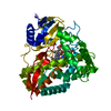
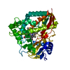
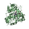
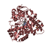
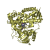
| Title | Crystal structure of Mycobacterium tuberculosis cytochrome P450 CYP125 in complex with econazole | ||||||
 Components Components | Cytochrome P450 CYP125 | ||||||
 Keywords Keywords | OXIDOREDUCTASE / econazole / cytochrome P450 / tuberculosis / monooxygenase / cholesterol / Heme / Iron / Metal-binding | ||||||
| Function / homology |  Function and homology information Function and homology informationcholest-4-en-3-one 26-monooxygenase [(25S)-3-oxocholest-4-en-26-oate forming] / cholest-4-en-3-one 26-monooxygenase activity / biological process involved in interaction with host / steroid hydroxylase activity / cholesterol catabolic process / iron ion binding / heme binding Similarity search - Function | ||||||
| Biological species |   Mycobacterium tuberculosis (bacteria) Mycobacterium tuberculosis (bacteria) | ||||||
| Method |  X-RAY DIFFRACTION / FOURIER SYNTHESIS / Resolution: 2.19 Å X-RAY DIFFRACTION / FOURIER SYNTHESIS / Resolution: 2.19 Å | ||||||
 Authors Authors | McLean, K.J. / Levy, C. / Munro, A.W. / Leys, D. | ||||||
 Citation Citation | Journal: J.Biol.Chem. / Year: 2009 Title: The Structure of Mycobacterium tuberculosis CYP125: molecular basis for cholesterol binding in a P450 needed for host infection. Authors: McLean, K.J. / Lafite, P. / Levy, C. / Cheesman, M.R. / Mast, N. / Pikuleva, I.A. / Leys, D. / Munro, A.W. | ||||||
| History |
|
- Structure visualization
| Structure viewer | Molecule: MolmilJmol/JSmol |
|---|
- Downloads & links
-Download
| PDBx/mmCIF format | 3iw2.cif.gz | 176.6 KB | Display | PDBx/mmCIF format |
|---|---|---|---|---|
| PDB format | pdb3iw2.ent.gz | 137.9 KB | Display | PDB format |
| PDBx/mmJSON format | 3iw2.json.gz | Tree view | PDBx/mmJSON format | |
| Others |  Other downloads Other downloads |
-Validation report
| Arichive directory | https://data.pdbj.org/pub/pdb/validation_reports/iw/3iw2ftp://data.pdbj.org/pub/pdb/validation_reports/iw/3iw2 | HTTPS FTP |
|---|
-Related structure data
| Related structure data | 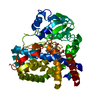 3ivySC 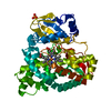 3iw0C 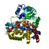 3iw1C S: Starting model for refinement C: citing same article ( |
|---|---|
| Similar structure data |


-Links
 PDBj
PDBj
- Assembly
| Deposited unit | 
| ||||||||
|---|---|---|---|---|---|---|---|---|---|
| 1 |
| ||||||||
| Unit cell |
|
-Components
| #1: Protein | Mass: 48491.492 Da / Num. of mol.: 1 Source method: isolated from a genetically manipulated source Details: Protein was produced in E.coli HMS174(DE3) (typically 15-20 L, grown in 2YT media) by IPTG (0.15 mM) induction in presence of the heme precursor delta aminolevulinic acid (delALA, 0.1 mM) at ...Details: Protein was produced in E.coli HMS174(DE3) (typically 15-20 L, grown in 2YT media) by IPTG (0.15 mM) induction in presence of the heme precursor delta aminolevulinic acid (delALA, 0.1 mM) at OD600 = 0.6, with temperature then reduced from 37 deg C to 23 deg C and culture continued for 24 hrs. Source: (gene. exp.) Mycobacterium tuberculosis (bacteria) / Strain: H37Rv / Gene: cyp125, MT3649, MTCY03C7.11, Rv3545c / Plasmid: pET15b / Production host: References: UniProt: P63709, UniProt: P9WPP1*PLUS, Oxidoreductases; Acting on paired donors, with incorporation or reduction of molecular oxygen |
|---|---|
| #2: Chemical | ChemComp-EKO /   Mass: 381.684 Da / Num. of mol.: 1 / Source method: obtained synthetically / Formula: C18H15Cl3N2O Mass: 381.684 Da / Num. of mol.: 1 / Source method: obtained synthetically / Formula: C18H15Cl3N2O |
| #3: Chemical | ChemComp-HEM /   Mass: 616.487 Da / Num. of mol.: 1 / Source method: obtained synthetically / Formula: C34H32FeN4O4 Mass: 616.487 Da / Num. of mol.: 1 / Source method: obtained synthetically / Formula: C34H32FeN4O4 |
| #4: Water | ChemComp-HOH /  Mass: 18.015 Da / Num. of mol.: 329 / Source method: isolated from a natural source / Formula: H2O Mass: 18.015 Da / Num. of mol.: 329 / Source method: isolated from a natural source / Formula: H2O |
-Experimental details
-Experiment
| Experiment | Method: X-RAY DIFFRACTION / Number of used crystals: 1 |
|---|
- Sample preparation
| Crystal | Density Matthews: 2.37 Å3/Da / Density % sol: 48 % |
|---|---|
| Crystal grow | Temperature: 277 K / Method: vapor diffusion, sitting drop / pH: 7.5 Details: Crystallization was refined to two different conditions, both consisting of MgCl2 with 0.1 M HEPES (either pH 7.0 or 7.5) and 20% PEG 6000 or 25% PEG 3350, respectively., VAPOR DIFFUSION, ...Details: Crystallization was refined to two different conditions, both consisting of MgCl2 with 0.1 M HEPES (either pH 7.0 or 7.5) and 20% PEG 6000 or 25% PEG 3350, respectively., VAPOR DIFFUSION, SITTING DROP, temperature 277K |
-Data collection
| Diffraction | Mean temperature: 100 K |
|---|---|
| Diffraction source | Source: ROTATING ANODE / Type: BRUKER AXS MICROSTAR / Wavelength: 1.5418 Å |
| Detector | Type: Bruker Platinum 135 / Detector: CCD / Date: Jul 1, 2008 / Details: mirrors |
| Radiation | Protocol: SINGLE WAVELENGTH / Monochromatic (M) / Laue (L): M / Scattering type: x-ray |
| Radiation wavelength | Wavelength: 1.5418 Å / Relative weight: 1 |
| Reflection | Resolution: 2.19→50 Å / Num. all: 21805 / Num. obs: 21805 / % possible obs: 94.22 % / Observed criterion σ(I): 0 / Rmerge(I) obs: 0.098 / Net I/σ(I): 8.7 |
| Reflection shell | Resolution: 2.19→2.3 Å |
- Processing
| Software |
| ||||||||||||||||||||||||||||||||||||||||||||||||||||||||||||||||||||||||||||||||||||||||||||||||||||||||||||||||||||||||||||||||||||||||||||||||||||||||||||||||||||||||||
|---|---|---|---|---|---|---|---|---|---|---|---|---|---|---|---|---|---|---|---|---|---|---|---|---|---|---|---|---|---|---|---|---|---|---|---|---|---|---|---|---|---|---|---|---|---|---|---|---|---|---|---|---|---|---|---|---|---|---|---|---|---|---|---|---|---|---|---|---|---|---|---|---|---|---|---|---|---|---|---|---|---|---|---|---|---|---|---|---|---|---|---|---|---|---|---|---|---|---|---|---|---|---|---|---|---|---|---|---|---|---|---|---|---|---|---|---|---|---|---|---|---|---|---|---|---|---|---|---|---|---|---|---|---|---|---|---|---|---|---|---|---|---|---|---|---|---|---|---|---|---|---|---|---|---|---|---|---|---|---|---|---|---|---|---|---|---|---|---|---|---|---|
| Refinement | Method to determine structure: FOURIER SYNTHESIS Starting model: PDB entry 3IVY Resolution: 2.19→49.63 Å / Cor.coef. Fo:Fc: 0.953 / Cor.coef. Fo:Fc free: 0.903 / SU B: 10.008 / SU ML: 0.139 / Cross valid method: THROUGHOUT / σ(F): 0 / ESU R Free: 0.218 / Stereochemistry target values: MAXIMUM LIKELIHOOD / Details: HYDROGENS HAVE BEEN ADDED IN THE RIDING POSITIONS
| ||||||||||||||||||||||||||||||||||||||||||||||||||||||||||||||||||||||||||||||||||||||||||||||||||||||||||||||||||||||||||||||||||||||||||||||||||||||||||||||||||||||||||
| Solvent computation | Ion probe radii: 0.8 Å / Shrinkage radii: 0.8 Å / VDW probe radii: 1.2 Å / Solvent model: MASK | ||||||||||||||||||||||||||||||||||||||||||||||||||||||||||||||||||||||||||||||||||||||||||||||||||||||||||||||||||||||||||||||||||||||||||||||||||||||||||||||||||||||||||
| Displacement parameters | Biso mean: 15.764 Å2
| ||||||||||||||||||||||||||||||||||||||||||||||||||||||||||||||||||||||||||||||||||||||||||||||||||||||||||||||||||||||||||||||||||||||||||||||||||||||||||||||||||||||||||
| Refinement step | Cycle: LAST / Resolution: 2.19→49.63 Å
| ||||||||||||||||||||||||||||||||||||||||||||||||||||||||||||||||||||||||||||||||||||||||||||||||||||||||||||||||||||||||||||||||||||||||||||||||||||||||||||||||||||||||||
| Refine LS restraints |
| ||||||||||||||||||||||||||||||||||||||||||||||||||||||||||||||||||||||||||||||||||||||||||||||||||||||||||||||||||||||||||||||||||||||||||||||||||||||||||||||||||||||||||
| LS refinement shell | Resolution: 2.19→2.249 Å / Total num. of bins used: 20
|
